The ZBED1 Antibody (CAB6792) is a high-quality antibody developed for reliable detection and analysis of target proteins. This antibody is produced in rabbits and exhibits high reactivity with human samples, making it ideal for use in Western blot applications. By specifically binding to the ZBED1 protein, researchers can accurately detect and analyze its presence in various cell types.ZBED1 is known to play a pivotal role in controlling gene expression related to cell proliferation, differentiation, and apoptosis. Its significance in these processes makes it a valuable target for research in fields such as developmental biology, cancer research, and regenerative medicine.
This antibody is validated for use in WB, IHC-P, IF/ICC, ELISA applications and has demonstrated reactivity against Human samples.
Product Name:
ZBED1 Antibody
SKU:
CAB6792
Size:
20μL, 100μL
Reactivity:
Human
Conjugate:
Unconjugated
Immunogen:
Recombinant protein (or fragment).This information is considered to be commercially sensitive.
Recommended starting concentration is 1 μg/mL. Please optimize the concentration based on your specific assay requirements.
Synonyms:
ALTE, DREF, TRAMP, hDREF, ZBED1
Positive Sample:
bewo
Cellular Localization:
Nucleus.
Calculated MW:
78kDa
Observed MW:
78kDa
This gene is located in the pseudoautosomal region 1 (PAR1) of X and Y chromosomes. It was earlier identified as a gene with similarity to Ac transposable elements, however, was found not to have transposase activity. Later studies show that this gene product is localized in the nucleus and functions as a transcription factor. It binds to DNA elements found in the promoter regions of several genes related to cell proliferation, such as histone H1, hence may have a role in regulating genes related to cell proliferation. Alternatively spliced transcript variants with different 5' untranslated region have been found for this gene.
Purification Method
Affinity purification
Gene ID
9189
RRID
AB_2767375
Buffer Information
Store at -20℃. Avoid freeze / thaw cycles. Buffer: PBS containing 50% glycerol, preserved with proclin300 or sodium azide, pH 7.3.
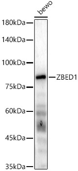
Western blot analysis of lysates from bewo cells, using ZBED1 Rabbit pAb (CAB6792) at 1:1000 dilution. Secondary antibody: HRP-conjugated Goat anti-Rabbit IgG (H+L) (CABS014) at 1:10000 dilution. Lysates/proteins: 25μg per lane. Blocking buffer: 3% nonfat dry milk in TBST. Detection: ECL Basic Kit (AbGn00020). Exposure time: 60s.
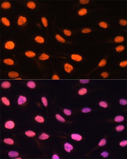
Immunofluorescence analysis of U-2 OS cells using ZBED1 Rabbit pAb (CAB6792) at dilution of 1:100. Secondary antibody: Cy3-conjugated Goat anti-Rabbit IgG (H+L) (CABS007) at 1:500 dilution. Blue: DAPI for nuclear staining.