The ZP3 Antibody (CAB13156) is a high-quality antibody developed for reliable detection and analysis of target proteins. This antibody, produced in rabbits, displays high reactivity towards human samples and is validated for use in Western blotting applications. By binding to the ZP3 protein, researchers can effectively detect and analyze this key molecule in various cell types, making it a valuable tool for studies in reproductive biology and fertility research.ZP3, a critical player in sperm-egg interaction and fertilization, is crucial for successful reproduction in mammals.
This antibody is validated for use in WB, ELISA, IF-P applications and has demonstrated reactivity against Human, Mouse, Rat samples.
Product Name:
ZP3 Antibody
SKU:
CAB13156
Size:
20μL, 100μL
Reactivity:
Human, Mouse, Rat
Conjugate:
Unconjugated
Immunogen:
Synthetic peptide. This information is considered to be commercially sensitive.
Recommended starting concentration is 1 μg/mL. Please optimize the concentration based on your specific assay requirements.
Synonyms:
ZPC, ZP3A, ZP3B, Zp-3, OOMD3, OZEMA3, ZP3
Positive Sample:
HeLa
Cellular Localization:
Cell Membrane, Secreted, Single-Pass Type I Membrane Protein, Extracellular Matrix, Extracellular Space.
Calculated MW:
47kDa
Observed MW:
47kDa
The zona pellucida is an extracellular matrix that surrounds the oocyte and early embryo. It is composed primarily of three or four glycoproteins with various functions during fertilization and preimplantation development. The protein encoded by this gene is a structural component of the zona pellucida and functions in primary binding and induction of the sperm acrosome reaction. The nascent protein contains a N-terminal signal peptide sequence, a conserved ZP domain, a C-terminal consensus furin cleavage site, and a transmembrane domain. It is hypothesized that furin cleavage results in release of the mature protein from the plasma membrane for subsequent incorporation into the zona pellucida matrix. However, the requirement for furin cleavage in this process remains controversial based on mouse studies. A variation in the last exon of this gene has previously served as the basis for an additional ZP3 locus; however, sequence and literature review reveals that there is only one full-length ZP3 locus in the human genome. Another locus encoding a bipartite transcript designated POMZP3 contains a duplication of the last four exons of ZP3, including the above described variation, and maps closely to this gene.
Purification Method
Affinity purification
Gene ID
7784
RRID
AB_2760006
Buffer Information
Store at -20℃. Avoid freeze / thaw cycles. Buffer: PBS with 0.01% thimerosal,50% glycerol,pH7.3.
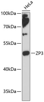
Western blot analysis of lysates from HeLa cells, using ZP3 Rabbit pAb (CAB13156) at 1:3000 dilution. Secondary antibody: HRP-conjugated Goat anti-Rabbit IgG (H+L) (CABS014) at 1:10000 dilution. Lysates/proteins: 25μg per lane. Blocking buffer: 3% nonfat dry milk in TBST. Detection: ECL Enhanced Kit (AbGn00021). Exposure time: 90s.
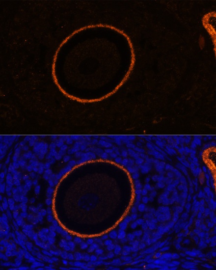
Immunofluorescence analysis of paraffin-embedded rat oophoroma using ZP3 Rabbit pAb (CAB13156) at dilution of 1:100. Secondary antibody: Cy3-conjugated Goat anti-Rabbit IgG (H+L) (CABS007) at 1:500 dilution. Blue: DAPI for nuclear staining.