The ACADM Monoclonal Antibody (CAB4567) is a high-quality antibody developed for reliable detection and analysis of target proteins. This antibody, produced in rabbits, exhibits high reactivity with human samples and has been validated for use in Western blot applications. It specifically binds to the ACADM protein, enabling accurate detection and analysis in a variety of cell types.ACADM plays a crucial role in breaking down fatty acids to produce energy, making it a key player in metabolic processes.
This antibody is validated for use in WB, IHC-P, IF/ICC, ELISA applications and has demonstrated reactivity against Human, Mouse, Rat samples.
Product Name:
ACADM Monoclonal Antibody
SKU:
CAB4567
Size:
20μL, 100μL
Reactivity:
Human, Mouse, Rat
Clone Number:
ARC1035
Conjugate:
Unconjugated
Immunogen:
Synthetic peptide. This information is considered to be commercially sensitive.
Recommended starting concentration is 1 μg/mL. Please optimize the concentration based on your specific assay requirements.
Synonyms:
MCAD, ACAD1, MCADH, ACADM
Positive Sample:
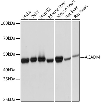
HeLa, 293T, Hep G2, Mouse liver, Mouse heart, Rat liver, Rat heart
Cellular Localization:
Mitochondrion Matrix.
Calculated MW:
47kDa
Observed MW:
47kDa
This gene encodes the medium-chain specific (C4 to C12 straight chain) acyl-Coenzyme A dehydrogenase. The homotetramer enzyme catalyzes the initial step of the mitochondrial fatty acid beta-oxidation pathway. Defects in this gene cause medium-chain acyl-CoA dehydrogenase deficiency, a disease characterized by hepatic dysfunction, fasting hypoglycemia, and encephalopathy, which can result in infantile death. Alternatively spliced transcript variants encoding different isoforms have been found for this gene.
Purification Method
Affinity purification
Gene ID
34
RRID
AB_2863298
Buffer Information
Store at -20℃. Avoid freeze / thaw cycles. Buffer: PBS containing 50% glycerol and 0.05% BSA, preserved with proclin300 or sodium azide, pH 7.3.
Western blot analysis of various lysates using ACADM Rabbit mAb (CAB4567) at 1:1000 dilution. Secondary antibody: HRP-conjugated Goat anti-Rabbit IgG (H+L) (CABS014) at 1:10000 dilution. Lysates/proteins: 25μg per lane. Blocking buffer: 3% nonfat dry milk in TBST. Detection: ECL Basic Kit (AbGn00020). Exposure time: 1s.
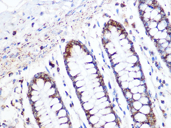
Immunohistochemistry analysis of paraffin-embedded Human colon using ACADM Rabbit mAb (CAB4567) at dilution of 1:100 (40x lens). Microwave antigen retrieval performed with 0.01M PBS Buffer (pH 7.2) prior to IHC staining.
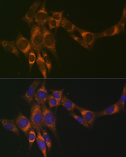
Immunofluorescence analysis of NIH/3T3 cells using ACADM Rabbit mAb (CAB4567) at dilution of 1:100 (40x lens). Secondary antibody: Cy3-conjugated Goat anti-Rabbit IgG (H+L) (CABS007) at 1:500 dilution. Blue: DAPI for nuclear staining.