The ACP1 Antibody (CAB1801) is a high-quality antibody developed for reliable detection and analysis of target proteins. This antibody, produced in rabbits, is highly specific to human ACP1 and has been validated for use in various applications, including Western blot analysis. By binding to the ACP1 enzyme, this antibody allows for precise detection and analysis in a variety of cell types, making it essential for studies in cell biology and disease research.
This antibody is validated for use in WB, ELISA applications and has demonstrated reactivity against Human, Mouse samples.
Product Name:
ACP1 Antibody
SKU:
CAB1801
Size:
20μL, 100μL
Reactivity:
Human, Mouse
Conjugate:
Unconjugated
Immunogen:
Recombinant protein (or fragment).This information is considered to be commercially sensitive.
Recommended starting concentration is 1 μg/mL. Please optimize the concentration based on your specific assay requirements.
Synonyms:
HAAP, LMWPTP, LMW-PTP, ACP1
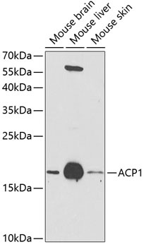
Positive Sample:
Mouse brain, Mouse liver, Mouse skin
Cellular Localization:
Cytoplasm.
Calculated MW:
18kDa
Observed MW:
18kDa
The product of this gene belongs to the phosphotyrosine protein phosphatase family of proteins. It functions as an acid phosphatase and a protein tyrosine phosphatase by hydrolyzing protein tyrosine phosphate to protein tyrosine and orthophosphate. This enzyme also hydrolyzes orthophosphoric monoesters to alcohol and orthophosphate. This gene is genetically polymorphic, and three common alleles segregating at the corresponding locus give rise to six phenotypes. Each allele appears to encode at least two electrophoretically different isozymes, Bf and Bs, which are produced in allele-specific ratios. Multiple alternatively spliced transcript variants encoding distinct isoforms have been identified for this gene.
Purification Method
Affinity purification
Gene ID
52
RRID
AB_2763840
Buffer Information
Store at -20℃. Avoid freeze / thaw cycles. Buffer: PBS containing 50% glycerol, preserved with proclin300 or sodium azide, pH 7.3.
Western blot analysis of various lysates using ACP1 Rabbit pAb (CAB1801) at 1:1000 dilution. Secondary antibody: HRP-conjugated Goat anti-Rabbit IgG (H+L) (CABS014) at 1:10000 dilution. Lysates/proteins: 25μg per lane. Blocking buffer: 3% nonfat dry milk in TBST. Detection: ECL Enhanced Kit (AbGn00021). Exposure time: 90s.