The AEBP1 Antibody (CAB16340) is a high-quality antibody developed for reliable detection and analysis of target proteins. This gene encodes a member of carboxypeptidase A protein family. The encoded protein may function as a transcriptional repressor and play a role in adipogenesis and smooth muscle cell differentiation. Studies in mice suggest that this gene functions in wound healing and abdominal wall development. Overexpression of this gene is associated with glioblastoma.
This antibody is validated for use in WB, IF/ICC, ELISA applications and has demonstrated reactivity against Human, Mouse, Rat samples.
Product Name:
AEBP1 Antibody
SKU:
CAB16340
Size:
100μL, 20μL
Reactivity:
Human, Mouse, Rat
Conjugate:
Unconjugated
Immunogen:
Recombinant protein (or fragment).This information is considered to be commercially sensitive.
Tested Applications:
WBIF/ICCELISA
Recommended Dilution:
WB
1:1000 - 1:5000
IF/ICC
1:50 - 1:100
ELISA
Recommended starting concentration is 1 μg/mL. Please optimize the concentration based on your specific assay requirements.
Synonyms:
ACLP, AEBP1
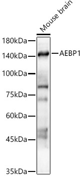
Positive Sample:
Mouse brain
Cellular Localization:
Cytoplasm, Nucleus, Secreted.
Calculated MW:
131kDa
Observed MW:
141kDa
This gene encodes a member of carboxypeptidase A protein family. The encoded protein may function as a transcriptional repressor and play a role in adipogenesis and smooth muscle cell differentiation. Studies in mice suggest that this gene functions in wound healing and abdominal wall development. Overexpression of this gene is associated with glioblastoma.
Purification Method
Affinity purification
Gene ID
165
RRID
AB_2768271
Buffer Information
Store at -20℃. Avoid freeze / thaw cycles. Buffer: PBS containing 50% glycerol, preserved with proclin300 or sodium azide, pH 7.3.
Western blot analysis of lysates from Mouse brain, using AEBP1 Rabbit pAb (CAB16340) at 1:2000 dilution. Secondary antibody: HRP-conjugated Goat anti-Rabbit IgG (H+L) (AS014) at 1:10000 dilution. Lysates/proteins: 25μg per lane. Blocking buffer: 3% nonfat dry milk in TBST. Detection: ECL Enhanced Kit (AbGn00021). Exposure time: 60s.
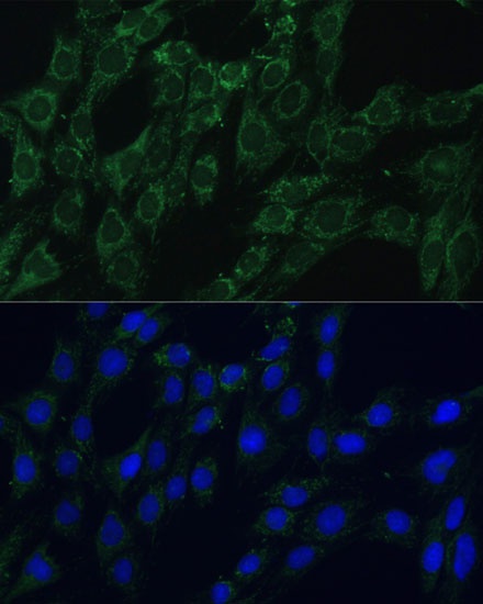
Immunofluorescence analysis of C6 cells using AEBP1 Rabbit pAb (CAB16340) at dilution of 1:100 (40x lens). Secondary antibody: Cy3-conjugated Goat anti-Rabbit IgG (H+L) (AS007) at 1:500 dilution. Blue: DAPI for nuclear staining.
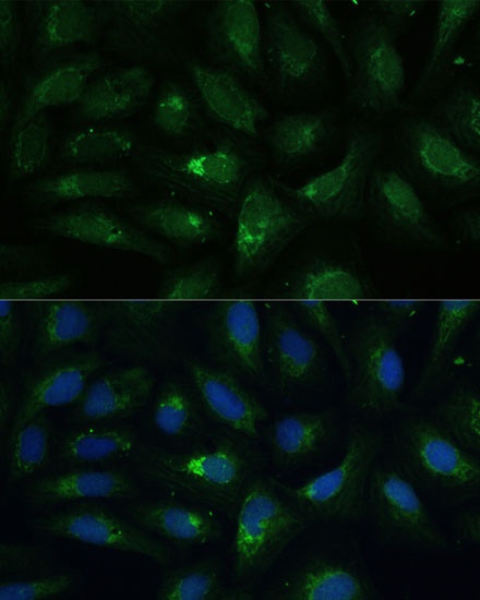
Immunofluorescence analysis of U-2 OS cells using AEBP1 Rabbit pAb (CAB16340) at dilution of 1:100 (40x lens). Secondary antibody: Cy3-conjugated Goat anti-Rabbit IgG (H+L) (AS007) at 1:500 dilution. Blue: DAPI for nuclear staining.