The AHR Antibody (CAB1451) is a high-quality antibody developed for reliable detection and analysis of target proteins. This antibody, produced in rabbits, has high specificity and sensitivity for detecting AHR protein in human samples, making it ideal for use in Western blot applications.The AHR protein is known to play a key role in the regulation of gene expression in response to environmental toxins and pollutants. It also has important functions in immune regulation, inflammation, and metabolic processes.
This antibody is validated for use in WB, IHC-P, IF/ICC, ELISA applications and has demonstrated reactivity against Human, Mouse, Rat samples.
Product Name:
AHR Antibody
SKU:
CAB1451
Size:
20μL, 100μL
Reactivity:
Human, Mouse, Rat
Conjugate:
Unconjugated
Immunogen:
Recombinant protein (or fragment).This information is considered to be commercially sensitive.
Recommended starting concentration is 1 μg/mL. Please optimize the concentration based on your specific assay requirements.
Synonyms:
RP85, bHLHe76, AHR
Positive Sample:
Hep G2, PC-3
Cellular Localization:
Cytoplasm, Nucleus.
Calculated MW:
96kDa
Observed MW:
100kDa
The protein encoded by this gene is a ligand-activated helix-loop-helix transcription factor involved in the regulation of biological responses to planar aromatic hydrocarbons. This receptor has been shown to regulate xenobiotic-metabolizing enzymes such as cytochrome P450. Before ligand binding, the encoded protein is sequestered in the cytoplasm; upon ligand binding, this protein moves to the nucleus and stimulates transcription of target genes.
Purification Method
Affinity purification
Gene ID
196
RRID
AB_2761384
Buffer Information
Store at -20℃. Avoid freeze / thaw cycles. Buffer: PBS containing 50% glycerol, preserved with proclin300 or sodium azide, pH 7.3.
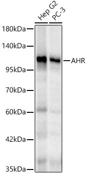
Western blot analysis of various lysates using AHR Rabbit pAb (CAB1451) at 1:2000 dilution. Secondary antibody: HRP-conjugated Goat anti-Rabbit IgG (H+L) (CABS014) at 1:10000 dilution. Lysates / proteins: 25 μg per lane. Blocking buffer: 3 % nonfat dry milk in TBST. Detection: ECL Basic Kit (AbGn00020). Exposure time: 10s.
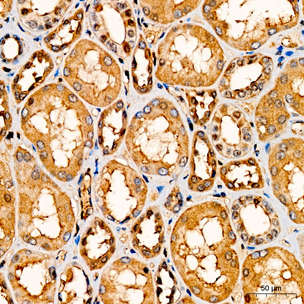
Immunohistochemistry analysis of paraffin-embedded Human kidney tissue using AHR Rabbit pAb (CAB1451) at a dilution of 1:200 (40x lens). High pressure antigen retrieval was performed with 0.01 M citrate buffer (pH 6.0) prior to IHC staining.
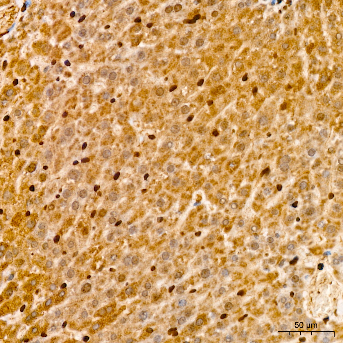
Immunohistochemistry analysis of paraffin-embedded Mouse liver tissue using AHR Rabbit pAb (CAB1451) at a dilution of 1:200 (40x lens). High pressure antigen retrieval was performed with 0.01 M citrate buffer (pH 6.0) prior to IHC staining.
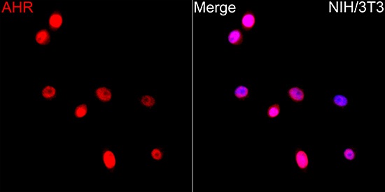
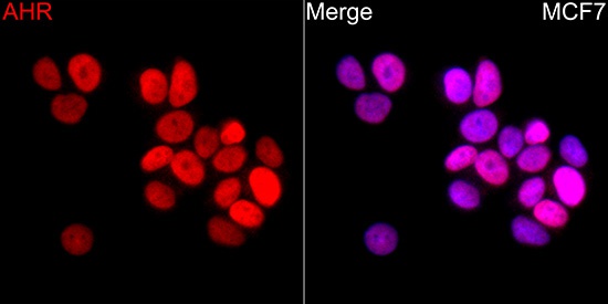
Immunofluorescence analysis of NIH/3T3 cells using AHR Rabbit pAb(CAB1451) at a dilution of 1:200 (40x lens). Secondary antibody:Cy3 Goat Anti-Rabbit IgG (H+L)(CABS007) at 1:500 dilution. Blue: DAPI for nuclear staining.
Western blot analysis of various lysates using AHR Rabbit pAb (CAB1451) at 1:2000 dilution. Secondary antibody: HRP-conjugated Goat anti-Rabbit IgG (H+L) (CABS014) at 1:10000 dilution. Lysates / proteins: 25 μg per lane. Blocking buffer: 3 % nonfat dry milk in TBST. Detection: ECL Basic Kit (AbGn00020). Exposure time: 10s.