The AK1 Antibody (CAB13460) is a high-quality antibody developed for reliable detection and analysis of target proteins. This antibody, produced in rabbits, exhibits a high level of reactivity with human samples and has been validated for use in Western blot applications. By specifically binding to the AK1 protein, researchers can easily detect and analyze AK1 expression in a variety of cell types, making it an essential component of studies focused on cellular energy regulation and metabolism.AK1, also known as adenylate kinase 1, plays a crucial role in maintaining cellular energy balance by facilitating the transfer of phosphate groups between nucleotides.
This antibody is validated for use in WB, IHC-P, ELISA applications and has demonstrated reactivity against Human, Mouse, Rat samples.
Product Name:
AK1 Antibody
SKU:
CAB13460
Size:
20μL, 100μL
Reactivity:
Human, Mouse, Rat
Conjugate:
Unconjugated
Immunogen:
Recombinant protein (or fragment).This information is considered to be commercially sensitive.
Recommended starting concentration is 1 μg/mL. Please optimize the concentration based on your specific assay requirements.
Synonyms:
HTL-S-58j, AK1
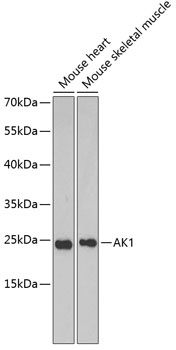
Positive Sample:
Mouse heart, Mouse skeletal muscle
Cellular Localization:
Cytoplasm.
Calculated MW:
22kDa
Observed MW:
22kDa
This gene encodes an adenylate kinase enzyme involved in energy metabolism and homeostasis of cellular adenine nucleotide ratios in different intracellular compartments. This gene is highly expressed in skeletal muscle, brain and erythrocytes. Certain mutations in this gene resulting in a functionally inadequate enzyme are associated with a rare genetic disorder causing nonspherocytic hemolytic anemia. Alternative splicing of this gene results in multiple transcript variants encoding different isoforms. This gene shares readthrough transcripts with the upstream ST6GALNAC6 gene.
Purification Method
Affinity purification
Gene ID
203
RRID
AB_2760321
Buffer Information
Store at -20℃. Avoid freeze / thaw cycles. Buffer: PBS containing 50% glycerol, preserved with proclin300 or sodium azide, pH 7.3.
Western blot analysis of various lysates using AK1 Rabbit pAb (CAB13460) at 1:1000 dilution. Secondary antibody: HRP-conjugated Goat anti-Rabbit IgG (H+L) (CABS014) at 1:10000 dilution. Lysates/proteins: 25μg per lane. Blocking buffer: 3% nonfat dry milk in TBST.
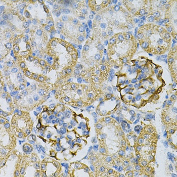
Immunohistochemistry analysis of paraffin-embedded Rat kidney using AK1 Rabbit pAb (CAB13460) at dilution of 1:100 (40x lens). Microwave antigen retrieval performed with 0.01M PBS Buffer (pH 7.2) prior to IHC staining.
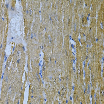
Immunohistochemistry analysis of paraffin-embedded Mouse heart using AK1 Rabbit pAb (CAB13460) at dilution of 1:100 (40x lens). Microwave antigen retrieval performed with 0.01M PBS Buffer (pH 7.2) prior to IHC staining.