The [KO Validated] ENO1 Antibody (CAB1033) is a high-quality antibody developed for reliable detection and analysis of target proteins. This gene encodes alpha-enolase, one of three enolase isoenzymes found in mammals. Each isoenzyme is a homodimer composed of 2 alpha, 2 gamma, or 2 beta subunits, and functions as a glycolytic enzyme. Alpha-enolase in addition, functions as a structural lens protein (tau-crystallin) in the monomeric form. Alternative splicing of this gene results in a shorter isoform that has been shown to bind to the c-myc promoter and function as a tumor suppressor. Several pseudogenes have been identified, including one on the long arm of chromosome 1. Alpha-enolase has also been identified as an autoantigen in Hashimoto encephalopathy. RRID AB_2757874 Gene ID 2023 Swiss Prot Synonym NNE; PPH; MPB1; ENO1L1; ENO1-IT1; HEL-S-17;ENO1
This antibody is validated for use in WB, IHC-P, IF/ICC, IP, ELISA applications and has demonstrated reactivity against Human, Mouse samples.
Product Name:
[KO Validated] ENO1 Antibody
SKU:
CAB1033
Size:
100μL, 20μL
Reactivity:
Human, Mouse
Clone Number:
-
Conjugate:
Unconjugated
Immunogen:
Recombinant protein (or fragment).This information is considered to be commercially sensitive.
Tested Applications:
WBIHC-PIF/ICCIPELISA
Recommended Dilution:
WB
1:1000 - 1:5000
IHC-P
1:50 - 1:200
IF
/
ICC
1:50 - 1:200
IP
0.5μg-4μg antibody for 200μg-400μg extracts of whole cells
ELISA
Recommended starting concentration is 1 μg/mL. Please optimize the concentration based on your specific assay requirements.
Synonyms:
NNE, PPH, MPB1, ENO1L1, ENO1-IT1, HEL-S-17, ENO1
Positive Sample:
HeLa, SH-SY5Y, MCF-7, Jurkat, NIH/3T3
Cellular Localization:
Cell Membrane, Cytoplasm, M Line, Nucleus, Myofibril, Sarcomere.
Calculated MW:
47kDa
Observed MW:
47kDa/
This gene encodes alpha-enolase, one of three enolase isoenzymes found in mammals. Each isoenzyme is a homodimer composed of 2 alpha, 2 gamma, or 2 beta subunits, and functions as a glycolytic enzyme. Alpha-enolase in addition, functions as a structural lens protein (tau-crystallin) in the monomeric form. Alternative splicing of this gene results in a shorter isoform that has been shown to bind to the c-myc promoter and function as a tumor suppressor. Several pseudogenes have been identified, including one on the long arm of chromosome 1. Alpha-enolase has also been identified as an autoantigen in Hashimoto encephalopathy. RRID AB_2757874 Gene ID 2023 Swiss Prot Synonym NNE; PPH; MPB1; ENO1L1; ENO1-IT1; HEL-S-17;ENO1
Purification Method:
Affinity purification
Gene ID:
2023
RRID:
AB_2757874
Buffer Information:
Store at -20℃. Avoid freeze / thaw cycles. Buffer: PBS with 0.09% sodium azide,50% glycerol,pH7.3.
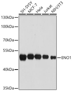
Western blot analysis of various lysates using ENO1 Rabbit pAb (CAB1033) at 1:3000 dilution incubated overnight at 4℃. Secondary antibody: HRP-conjugated Goat anti-Rabbit IgG (H+L) (AS014) at 1:10000 dilution. Lysates/proteins: 25 μg per lane. Blocking buffer: 3% nonfat dry milk in TBST. Detection: ECL Basic Kit (AbGn00020) Exposure time: 1 s.
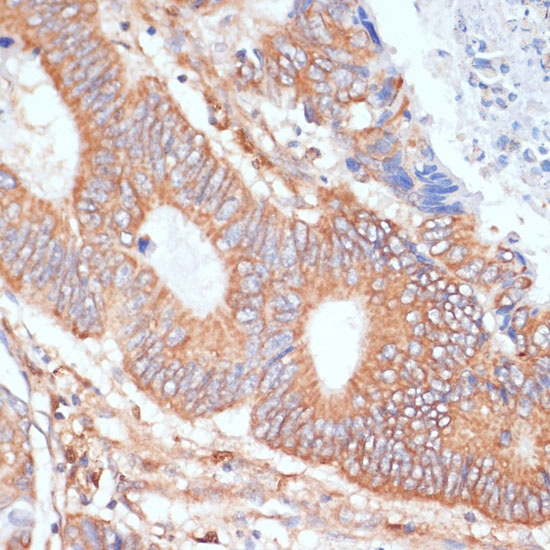
Immunohistochemistry analysis of paraffin-embedded Human colon carcinoma using ENO1 Rabbit pAb (CAB1033) at dilution of 1:100 (40x lens). Microwave antigen retrieval performed with 0.01M PBS Buffer (pH 7.2) prior to IHC staining.
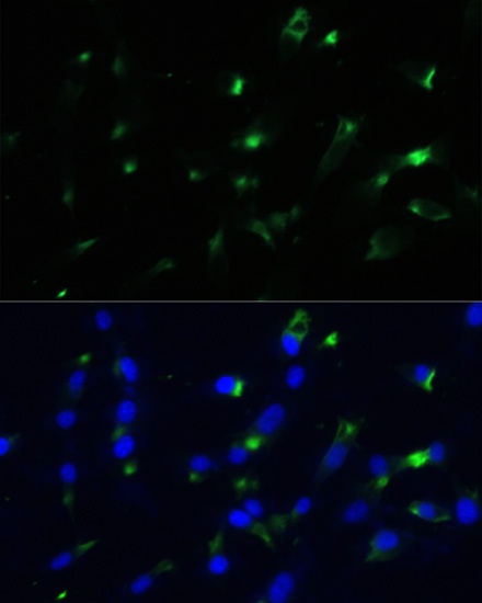
Immunofluorescence analysis of NIH-3T3 cells using ENO1 Rabbit pAb (CAB1033) at dilution of 1:100. Secondary antibody: Cy3-conjugated Goat anti-Rabbit IgG (H+L) (AS007) at 1:500 dilution. Blue: DAPI for nuclear staining.
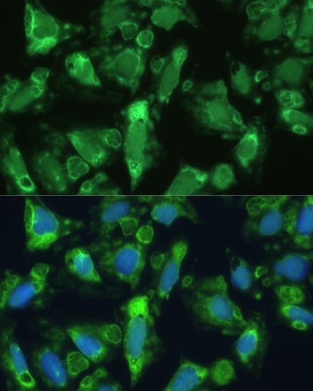
Immunofluorescence analysis of U-2 OS cells using ENO1 Rabbit pAb (CAB1033) at dilution of 1:100. Secondary antibody: Cy3-conjugated Goat anti-Rabbit IgG (H+L) (AS007) at 1:500 dilution. Blue: DAPI for nuclear staining.
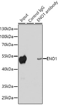
Immunoprecipitation analysis of 200 μg extracts of HeLa cells using 1 μg ENO1 Rabbit pAb (CAB1033). Western blot was performed from the immunoprecipitate using ENO1 antibody (CAB1033) at a dilution of 1:1000.