The AMD1 Antibody (CAB8537) is a high-quality antibody developed for reliable detection and analysis of target proteins. This polyclonal antibody, produced in rabbits, exhibits high specificity and sensitivity for detecting AMD1 in a variety of samples, particularly in Western blotting applications.AMD1, also known as adenosylmethionine decarboxylase 1, plays a crucial role in polyamine biosynthesis, which is essential for cell growth and survival. Dysregulation of AMD1 has been implicated in various diseases, including cancer and metabolic disorders.
This antibody is validated for use in WB, ELISA applications and has demonstrated reactivity against Human, Mouse, Rat samples.
Product Name:
AMD1 Antibody
SKU:
CAB8537
Size:
20μL, 100μL
Reactivity:
Human, Mouse, Rat
Conjugate:
Unconjugated
Immunogen:
Recombinant protein (or fragment).This information is considered to be commercially sensitive.
Recommended starting concentration is 1 μg/mL. Please optimize the concentration based on your specific assay requirements.
Synonyms:
AMD, SAMDC, ADOMETDC, AMD1
Positive Sample:
293T, Mouse skeletal muscle, Rat testis
Cellular Localization:
Cytosol.
Calculated MW:
38kDa
Observed MW:
32kDa/42kDa
This gene encodes an important intermediate enzyme in polyamine biosynthesis. The polyamines spermine, spermidine, and putrescine are low-molecular-weight aliphatic amines essential for cellular proliferation and tumor promotion. Multiple alternatively spliced transcript variants have been identified. Pseudogenes of this gene are found on chromosomes 5, 6, 10, X and Y.
Purification Method
Affinity purification
Gene ID
262
RRID
AB_2768346
Buffer Information
Store at -20℃. Avoid freeze / thaw cycles. Buffer: PBS containing 50% glycerol, preserved with proclin300 or sodium azide, pH 7.3.
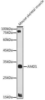
Western blot analysis of lysates from Mouse skeletal muscle, using AMD1 Rabbit pAb (CAB8537) at 1:1000 dilution. Secondary antibody: HRP-conjugated Goat anti-Rabbit IgG (H+L) (CABS014) at 1:10000 dilution. Lysates/proteins: 25μg per lane. Blocking buffer: 3% nonfat dry milk in TBST. Detection: ECL Basic Kit (AbGn00020). Exposure time: 60s.
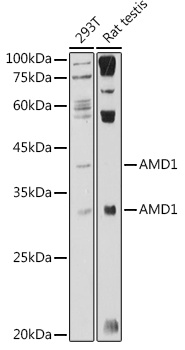
Western blot analysis of various lysates using AMD1 Rabbit pAb (CAB8537) at 1:1000 dilution. Secondary antibody: HRP-conjugated Goat anti-Rabbit IgG (H+L) (CABS014) at 1:10000 dilution. Lysates/proteins: 25μg per lane. Blocking buffer: 3% nonfat dry milk in TBST. Detection: ECL Basic Kit (AbGn00020). Exposure time: 180s.