The Androgen Receptor Monoclonal Antibody (CAB19611) is a high-quality antibody developed for reliable detection and analysis of target proteins. This antibody, generated in rabbits, exhibits high specificity and sensitivity in detecting the androgen receptor in human samples, providing reliable results for Western blotting and immunohistochemistry applications.The androgen receptor is a nuclear hormone receptor that plays a key role in the development and progression of prostate cancer, making it a target of interest for therapeutic interventions.
This antibody is validated for use in WB, IHC-P, IP, ELISA, IF-P, mIHC applications and has demonstrated reactivity against Human, Mouse, Rat samples.
Product Name:
Androgen Receptor Monoclonal Antibody
SKU:
CAB19611
Size:
20μL, 100μL
Reactivity:
Human, Mouse, Rat
Clone Number:
ARC0090
Conjugate:
Unconjugated
Immunogen:
Synthetic peptide. This information is considered to be commercially sensitive.
The androgen receptor gene is more than 90 kb long and codes for a protein that has 3 major functional domains: the N-terminal domain, DNA-binding domain, and androgen-binding domain. The protein functions as a steroid-hormone activated transcription factor. Upon binding the hormone ligand, the receptor dissociates from accessory proteins, translocates into the nucleus, dimerizes, and then stimulates transcription of androgen responsive genes. This gene contains 2 polymorphic trinucleotide repeat segments that encode polyglutamine and polyglycine tracts in the N-terminal transactivation domain of its protein. Expansion of the polyglutamine tract from the normal 9-34 repeats to the pathogenic 38-62 repeats causes spinal bulbar muscular atrophy (SBMA, also known as Kennedy's disease). Mutations in this gene are also associated with complete androgen insensitivity (CAIS). Alternative splicing results in multiple transcript variants encoding different isoforms.
Purification Method
Affinity purification
Gene ID
367
RRID
AB_2862699
Buffer Information
Store at -20℃. Avoid freeze / thaw cycles. Buffer: PBS containing 50% glycerol and 0.05% BSA, preserved with proclin300 or sodium azide, pH 7.3.
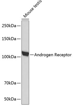
Western blot analysis of lysates from mouse testis, using Androgen Receptor Rabbit mAb (CAB19611) at 1:1000 dilution. Secondary antibody: HRP-conjugated Goat anti-Rabbit IgG (H+L) (CABS014) at 1:10000 dilution. Lysates/proteins: 25μg per lane. Blocking buffer: 3% nonfat dry milk in TBST. Detection: ECL Basic Kit (AbGn00020). Exposure time: 90s.
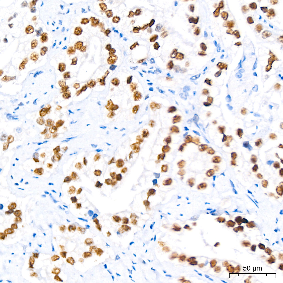
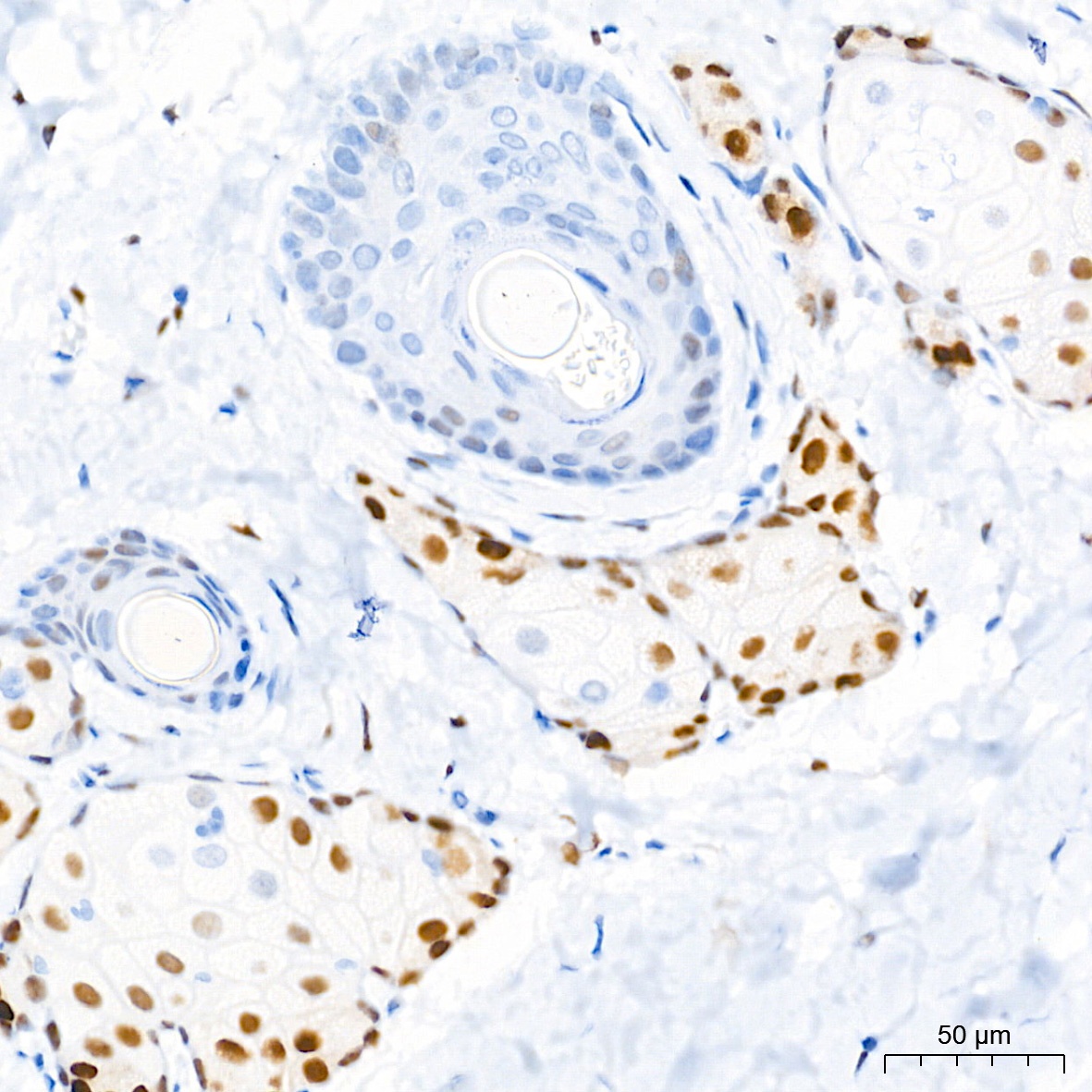
Immunohistochemistry analysis of paraffin-embedded Human prostate cancer tissue using Androgen Receptor Rabbit mAb (CAB19611) at dilution of 1:500 (40x lens). High pressure antigen retrieval performed with 0.01M Citrate Buffer (pH 6.0) prior to IHC staining.
Immunohistochemistry analysis of paraffin-embedded Human breast cancer tissue using Androgen Receptor Rabbit mAb (CAB19611) at a dilution of 1:500 (40x lens). High pressure antigen retrieval performed with 0.01M Tris-EDTA Buffer (pH 9.0) prior to IHC staining.
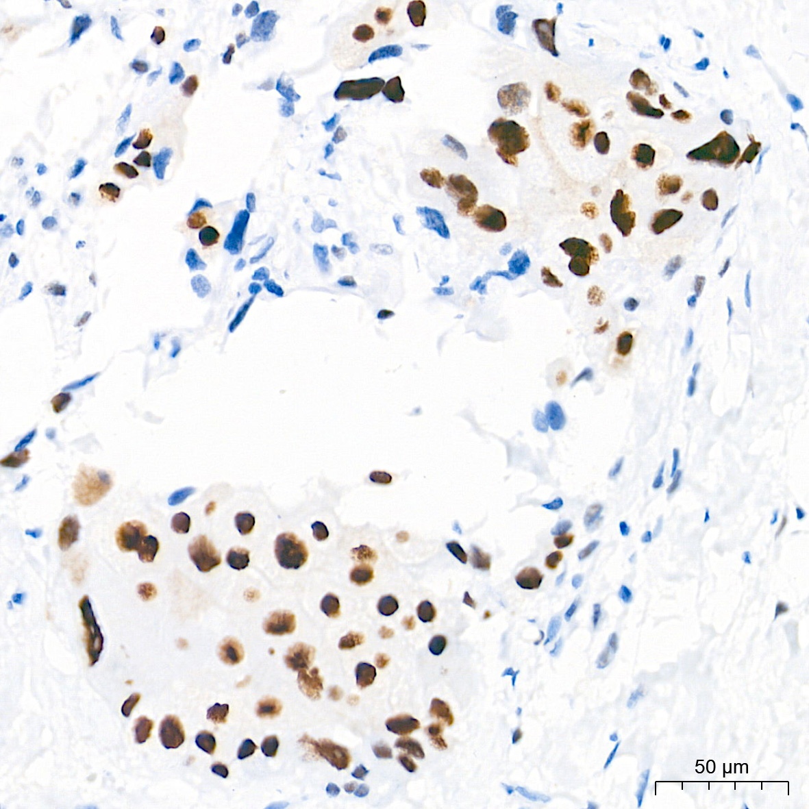
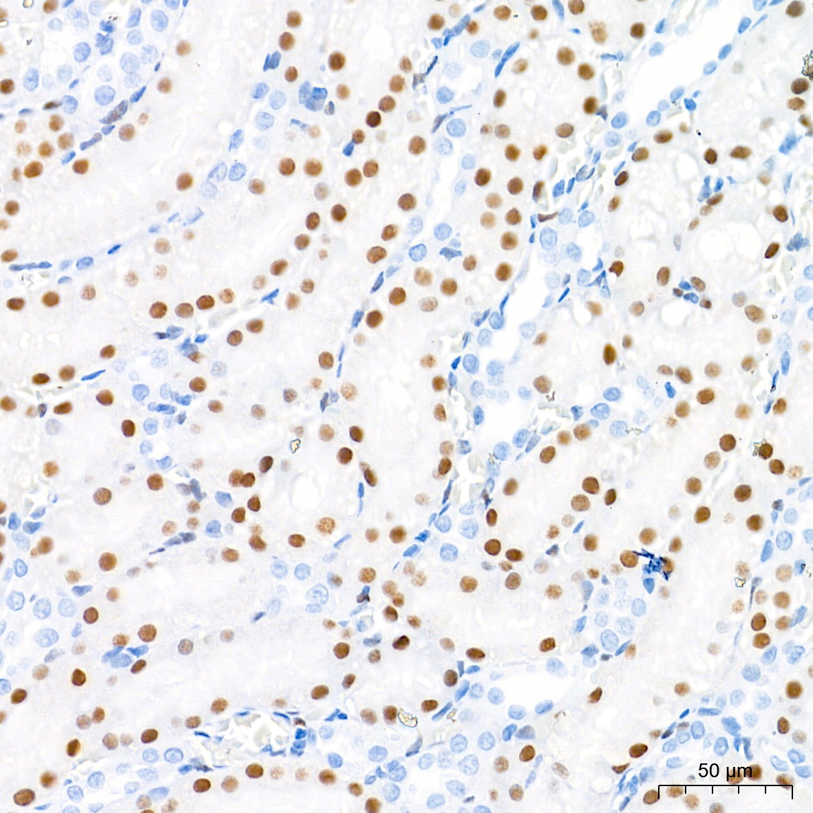
Immunohistochemistry analysis of paraffin-embedded Human testis tissue using Androgen Receptor Rabbit mAb (CAB19611) at a dilution of 1:500 (40x lens). High pressure antigen retrieval performed with 0.01M Tris-EDTA Buffer (pH 9.0) prior to IHC staining.
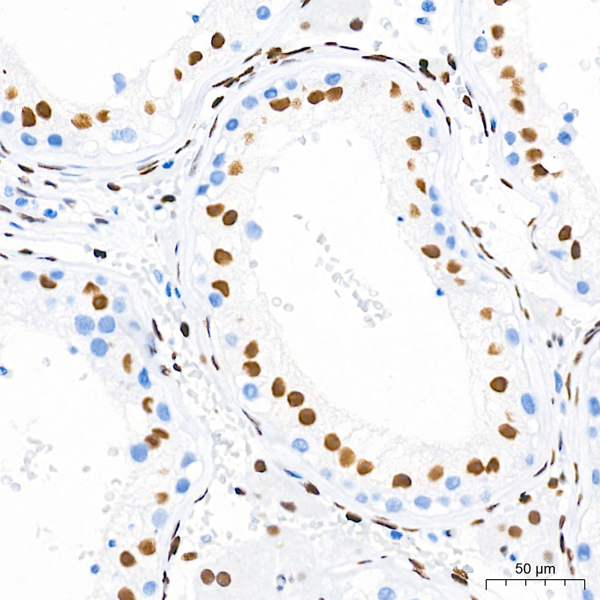
Immunohistochemistry analysis of paraffin-embedded Rat kidney tissue using Androgen Receptor Rabbit mAb (CAB19611) at a dilution of 1:500 (40x lens). High pressure antigen retrieval performed with 0.01M Tris-EDTA Buffer (pH 9.0) prior to IHC staining.
Immunohistochemistry analysis of paraffin-embedded Rat skin tissue using Androgen Receptor Rabbit mAb (CAB19611) at a dilution of 1:500 (40x lens). High pressure antigen retrieval performed with 0.01M Tris-EDTA Buffer (pH 9.0) prior to IHC staining.
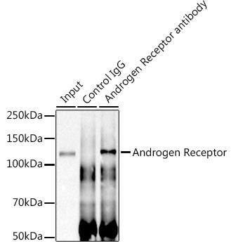
Immunoprecipitation analysis of 600 μg extracts of Mouse testis cells using 3 μg Androgen Receptor antibody (CAB19611). Western blot was performed from the immunoprecipitate using Androgen Receptor antibody (CAB19611) at a dilution of 1:1000.
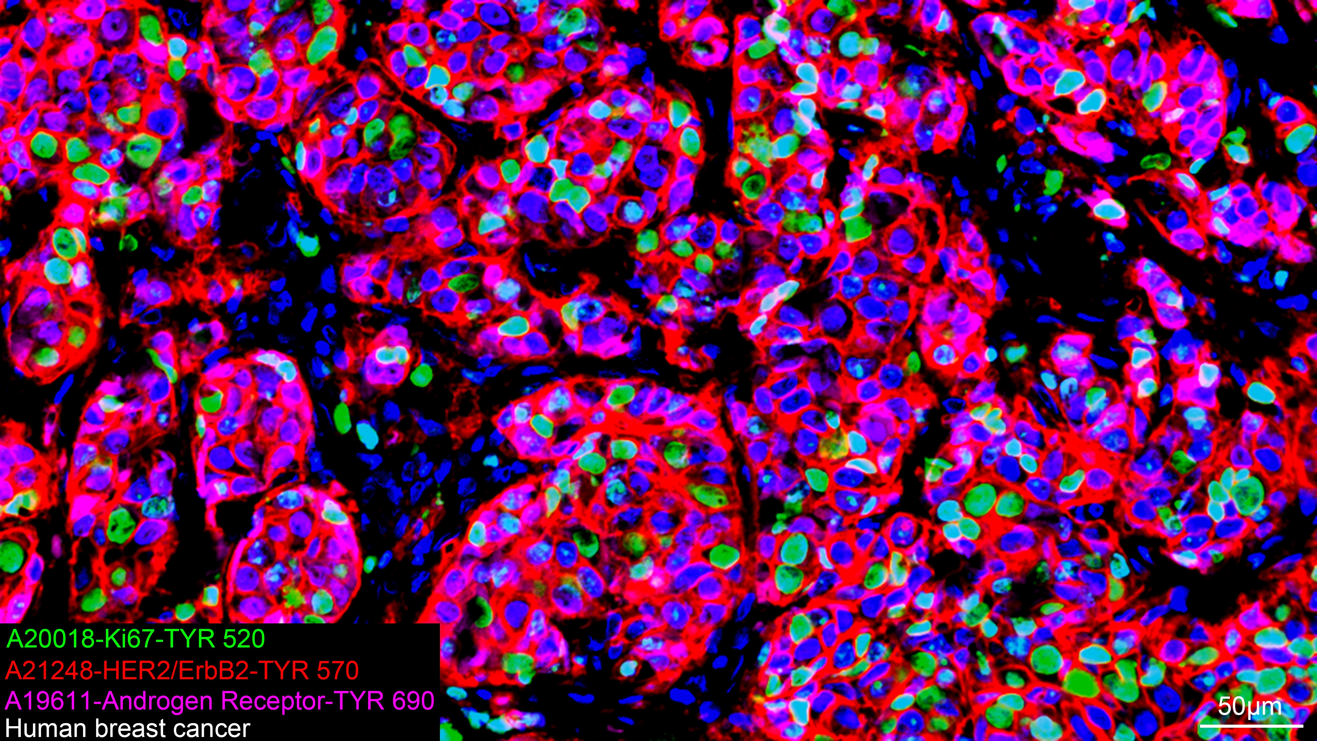
The multiplex IHC analysis on paraffin-embedded Human breast cancer tissue using the following specific primary antibodies and tyramide signal amplification (TSA) reagents (RK05903) : Ki67 Rabbit mAb (CAB20018, 1:500) with TSA-TYR-520 (Green), HER2/ErbB2 Rabbit mAb (CAB21248, 1:200) with TSA-TYR-570 (Red), and Androgen Receptor Rabbit mAb (CAB19611, 1:400) with TSA-TYR-690 (Magenta). DAPI (Blue) was used for nuclear staining. Prior to multiplex IHC staining, high-pressure antigen retrieval was performed using 0.01M citrate buffer at pH 6.0. The analysis was completed using a 20x objective lens.