The ALOX5 Monoclonal Antibody (CAB2877) is a high-quality antibody developed for reliable detection and analysis of target proteins. This polyclonal antibody, produced in rabbits, is highly specific to human ALOX5 and is validated for use in Western blot applications, allowing for accurate detection and analysis of the enzyme in various cell types.ALOX5 is an important enzyme in the 5-lipoxygenase pathway, which is responsible for the synthesis of leukotrienes that are potent pro-inflammatory mediators in conditions such as asthma, atherosclerosis, and inflammatory bowel disease.
This antibody is validated for use in WB, IHC-P, IF/ICC, ELISA applications and has demonstrated reactivity against Human, Mouse, Rat samples.
Product Name:
ALOX5 Monoclonal Antibody
SKU:
CAB2877
Size:
20μL, 100μL
Reactivity:
Human, Mouse, Rat
Clone Number:
ARC1926
Conjugate:
Unconjugated
Immunogen:
Synthetic peptide. This information is considered to be commercially sensitive.
This gene encodes a member of the lipoxygenase gene family and plays a dual role in the synthesis of leukotrienes from arachidonic acid. The encoded protein, which is expressed specifically in bone marrow-derived cells, catalyzes the conversion of arachidonic acid to 5(S)-hydroperoxy-6-trans-8,11,14-cis-eicosatetraenoic acid, and further to the allylic epoxide 5(S)-trans-7,9-trans-11,14-cis-eicosatetrenoic acid (leukotriene A4). Leukotrienes are important mediators of a number of inflammatory and allergic conditions. Mutations in the promoter region of this gene lead to a diminished response to antileukotriene drugs used in the treatment of asthma and may also be associated with atherosclerosis and several cancers. Alternatively spliced transcript variants encoding different isoforms have been found for this gene.
Purification Method
Affinity purification
Gene ID
240
RRID
AB_2863022
Buffer Information
Store at -20℃. Avoid freeze / thaw cycles. Buffer: PBS containing 50% glycerol and 0.05% BSA, preserved with proclin300 or sodium azide, pH 7.3.
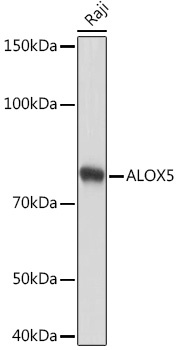
Western blot analysis of lysates from Raji cells, using ALOX5 Rabbit mAb (CAB2877) at 1:1000 dilution. Secondary antibody: HRP-conjugated Goat anti-Rabbit IgG (H+L) (CABS014) at 1:10000 dilution. Lysates/proteins: 25μg per lane. Blocking buffer: 3% nonfat dry milk in TBST. Detection: ECL Basic Kit (AbGn00020). Exposure time: 10s.
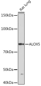
Western blot analysis of lysates from Rat lung, using ALOX5 Rabbit mAb (CAB2877) at 1:1000 dilution. Secondary antibody: HRP-conjugated Goat anti-Rabbit IgG (H+L) (CABS014) at 1:10000 dilution. Lysates/proteins: 25μg per lane. Blocking buffer: 3% nonfat dry milk in TBST. Detection: ECL Basic Kit (AbGn00020). Exposure time: 3min.
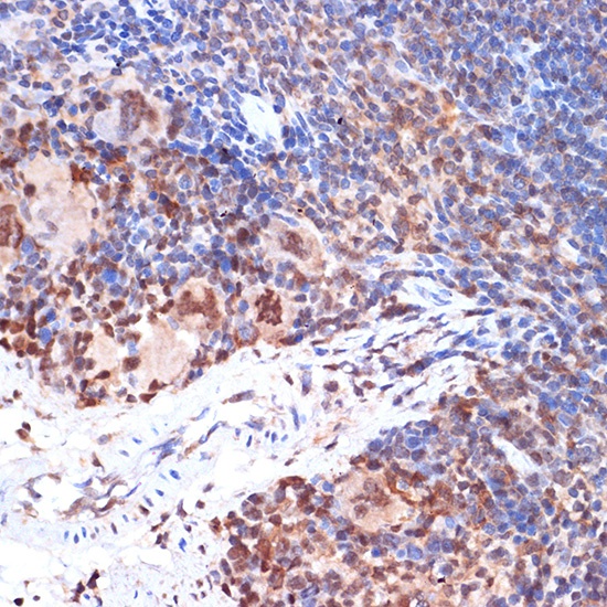
Immunohistochemistry analysis of paraffin-embedded Mouse spleen tissue using ALOX5 Rabbit mAb (CAB2877) at a dilution of 1:100 (40x lens). Microwave antigen retrieval performed with 0.01M Tris-EDTA Buffer (pH 9.0) prior to IHC staining.
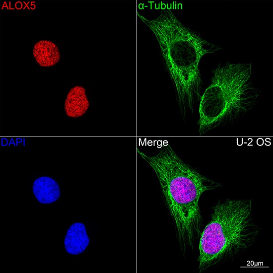
Confocal imaging of U-2 OS cells using ALOX5 Rabbit mAb (CAB2877,dilution 1:100)(Red). The cells were counterstained with α-Tubulin Mouse mAb (AC012,dilution 1:400) (Green). DAPI was used for nuclear staining (blue). Objective: 100x.