The CD3G Monoclonal Antibody (CAB4085) is a high-quality antibody developed for reliable detection and analysis of target proteins. This antibody, generated in rabbits, exhibits high reactivity with human samples and has been validated for use in Western blot applications. By binding to the CD3G protein, this antibody facilitates the detection and analysis of CD3G in various cell types, making it an ideal choice for studies in immunology and cancer research.CD3G is a crucial protein involved in T-cell signaling and activation, playing a pivotal role in the adaptive immune response. Dysregulation of CD3G expression or function can have profound implications for immune function and has been implicated in various diseases, including autoimmune disorders and cancer.
This antibody is validated for use in WB, IHC-P, IF/ICC, ELISA applications and has demonstrated reactivity against Human, Mouse samples.
Product Name:
CD3G Monoclonal Antibody
SKU:
CAB4085
Size:
20μL, 100μL
Reactivity:
Human, Mouse
Clone Number:
ARC2105
Conjugate:
Unconjugated
Immunogen:
Synthetic peptide. This information is considered to be commercially sensitive.
Recommended starting concentration is 1 μg/mL. Please optimize the concentration based on your specific assay requirements.
Synonyms:
T3G, IMD17, CD3GAMMA, CD3-GAMMA, CD3G
Positive Sample:
Jurkat
Cellular Localization:
Cell Membrane.
Calculated MW:
20kDa
Observed MW:
20kDa/25kDa
The protein encoded by this gene is the CD3-gamma polypeptide, which together with CD3-epsilon, -delta and -zeta, and the T-cell receptor alpha/beta and gamma/delta heterodimers, forms the T-cell receptor-CD3 complex. This complex plays an important role in coupling antigen recognition to several intracellular signal-transduction pathways. The genes encoding the epsilon, gamma and delta polypeptides are located in the same cluster on chromosome 11. Defects in this gene are associated with T cell immunodeficiency.
Purification Method
Affinity purification
Gene ID
917
Buffer Information
Store at -20℃. Avoid freeze / thaw cycles. Buffer: PBS containing 50% glycerol and 0.05% BSA, preserved with proclin300 or sodium azide, pH 7.3.
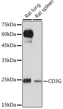
Western blot analysis of various lysates using CD3G Rabbit mAb (CAB4085) at 1:1000 dilution. Secondary antibody: HRP-conjugated Goat anti-Rabbit IgG (H+L) (CABS014) at 1:10000 dilution. Lysates/proteins: 25μg per lane. Blocking buffer: 3% nonfat dry milk in TBST. Detection: ECL Basic Kit (AbGn00020). Exposure time: 120s.
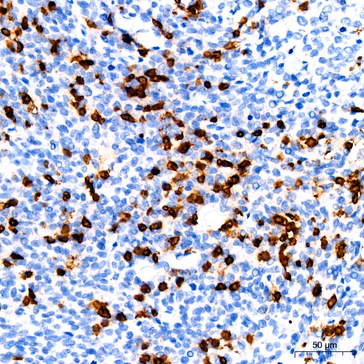
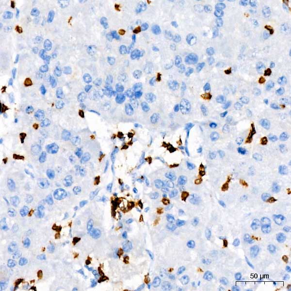
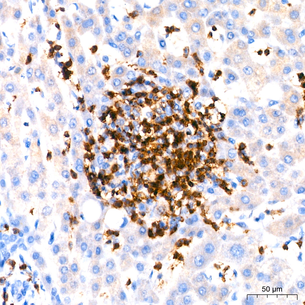
Immunohistochemistry analysis of paraffin-embedded Human cervix cancer tissue using CD3G Rabbit mAb (CAB4085) at a dilution of 1:100 (40x lens). High pressure antigen retrieval performed with 0.01M Citrate Buffer (pH 6.0) prior to IHC staining.
Immunohistochemistry analysis of paraffin-embedded Human liver cancer tissue using CD3G Rabbit mAb (CAB4085) at a dilution of 1:100 (40x lens). High pressure antigen retrieval performed with 0.01M Citrate Buffer (pH 6.0) prior to IHC staining.
Immunohistochemistry analysis of paraffin-embedded Human liver tissue using CD3G Rabbit mAb (CAB4085) at a dilution of 1:100 (40x lens). High pressure antigen retrieval performed with 0.01M Citrate Buffer (pH 6.0) prior to IHC staining.
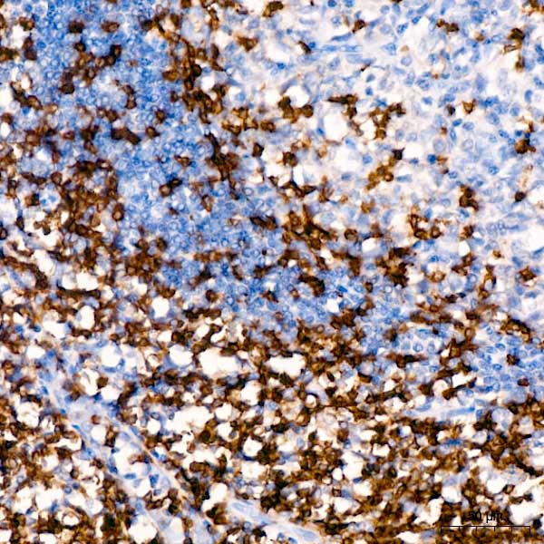
Immunohistochemistry analysis of paraffin-embedded Human tonsil tissue using CD3G Rabbit mAb (CAB4085) at a dilution of 1:100 (40x lens). High pressure antigen retrieval performed with 0.01M Citrate Buffer (pH 6.0) prior to IHC staining.
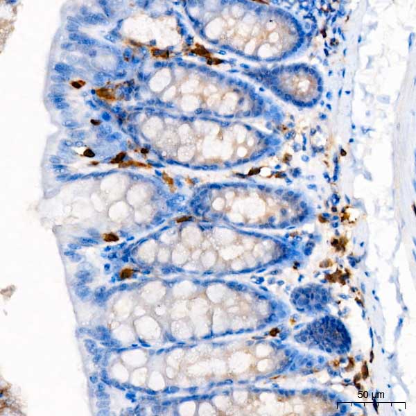
Immunohistochemistry analysis of paraffin-embedded Mouse colon tissue using CD3G Rabbit mAb (CAB4085) at a dilution of 1:100 (40x lens). High pressure antigen retrieval performed with 0.01M Citrate Buffer (pH 6.0) prior to IHC staining.
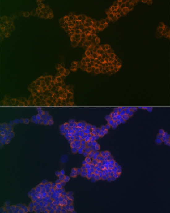
Immunofluorescence analysis of Jurkat cells using CD3G Rabbit mAb (CAB4085) at dilution of 1:100 (40x lens). Secondary antibody: Cy3-conjugated Goat anti-Rabbit IgG (H+L) (CABS007) at 1:500 dilution. Blue: DAPI for nuclear staining.