The Chromogranin A Monoclonal Antibody (CAB9576) is a high-quality antibody developed for reliable detection and analysis of target proteins. This polyclonal antibody, generated in rabbits, has high reactivity with human samples and is validated for use in various applications such as immunohistochemistry, immunofluorescence, and ELISA.Chromogranin A is a key marker for neuroendocrine tumors and can be used to diagnose and monitor patients with these types of cancers. By using this antibody, researchers can detect and analyze Chromogranin A expression in tissues and cells, allowing for a better understanding of its role in cancer biology and potential therapeutic targets.
This antibody is validated for use in IHC-P, ELISA, IF-P applications and has demonstrated reactivity against Human, Mouse, Rat samples.
Product Name:
Chromogranin A Monoclonal Antibody
SKU:
CAB9576
Size:
20μL, 100μL
Reactivity:
Human, Mouse, Rat
Clone Number:
ARC1643
Conjugate:
Unconjugated
Immunogen:
Synthetic peptide. This information is considered to be commercially sensitive.
Recommended starting concentration is 1 μg/mL. Please optimize the concentration based on your specific assay requirements.
Synonyms:
CGA, PHE5, PHES, Chromogranin A
Cellular Localization:
Chromaffin Granule, Extracellular Region, Extracellular Space, Perinuclear Region Of Cytoplasm.
Calculated MW:
51kDa
Observed MW:
Refertofigures
The protein encoded by this gene is a member of the chromogranin/secretogranin family of neuroendocrine secretory proteins. It is found in secretory vesicles of neurons and endocrine cells. This gene product is a precursor to three biologically active peptides; vasostatin, pancreastatin, and parastatin. These peptides act as autocrine or paracrine negative modulators of the neuroendocrine system. Two other peptides, catestatin and chromofungin, have antimicrobial activity and antifungal activity, respectively. Two transcript variants encoding different isoforms have been found for this gene.
Purification Method
Affinity purification
Gene ID
1113
Buffer Information
Store at -20℃. Avoid freeze / thaw cycles. Buffer: PBS containing 50% glycerol and 0.05% BSA, preserved with proclin300 or sodium azide, pH 7.3.
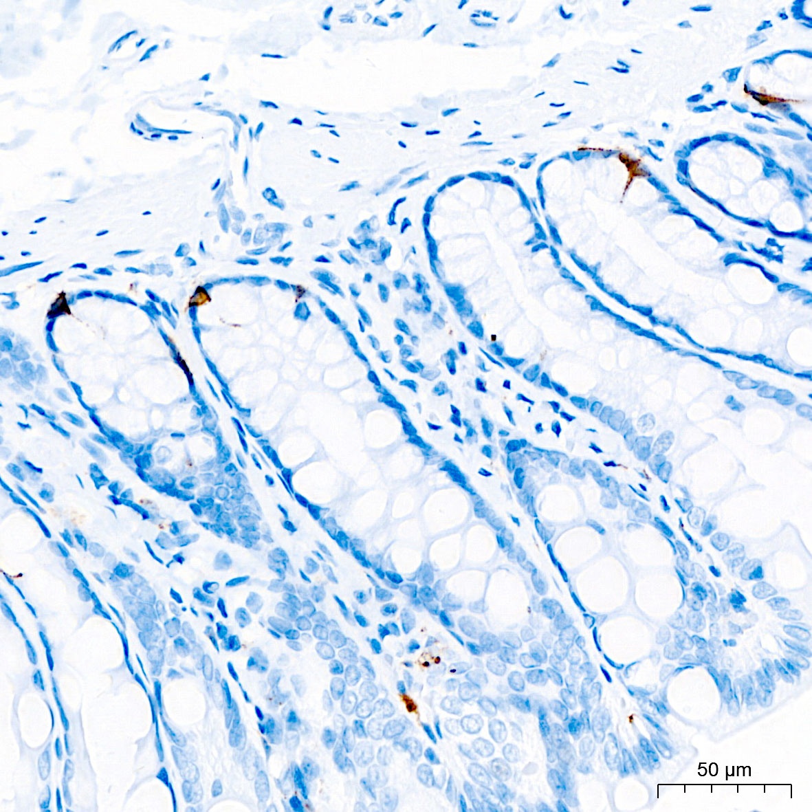
Immunohistochemistry analysis of paraffin-embedded Rat colon tissue using Chromogranin A Rabbit mAb (CAB9576) at a dilution of 1:4000 (40x lens). High pressure antigen retrieval performed with 0.01M Citrate Buffer (pH 6.0) prior to IHC staining.
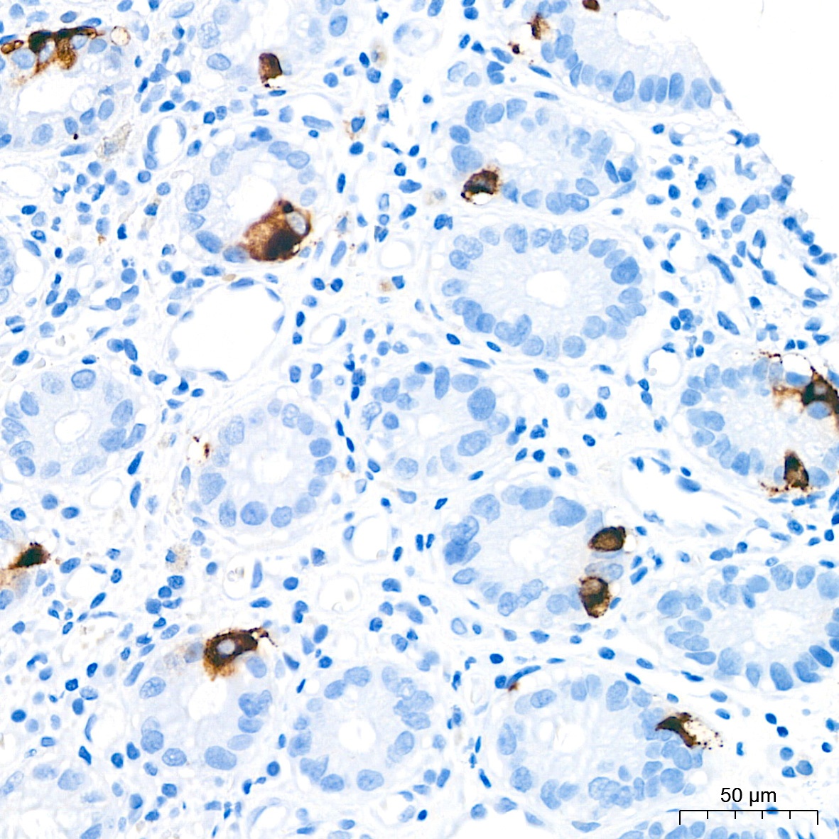
Immunohistochemistry analysis of paraffin-embedded Human colon tissue using Chromogranin A Rabbit mAb (CAB9576) at a dilution of 1:4000 (40x lens). High pressure antigen retrieval performed with 0.01M Citrate Buffer (pH 6.0) prior to IHC staining.
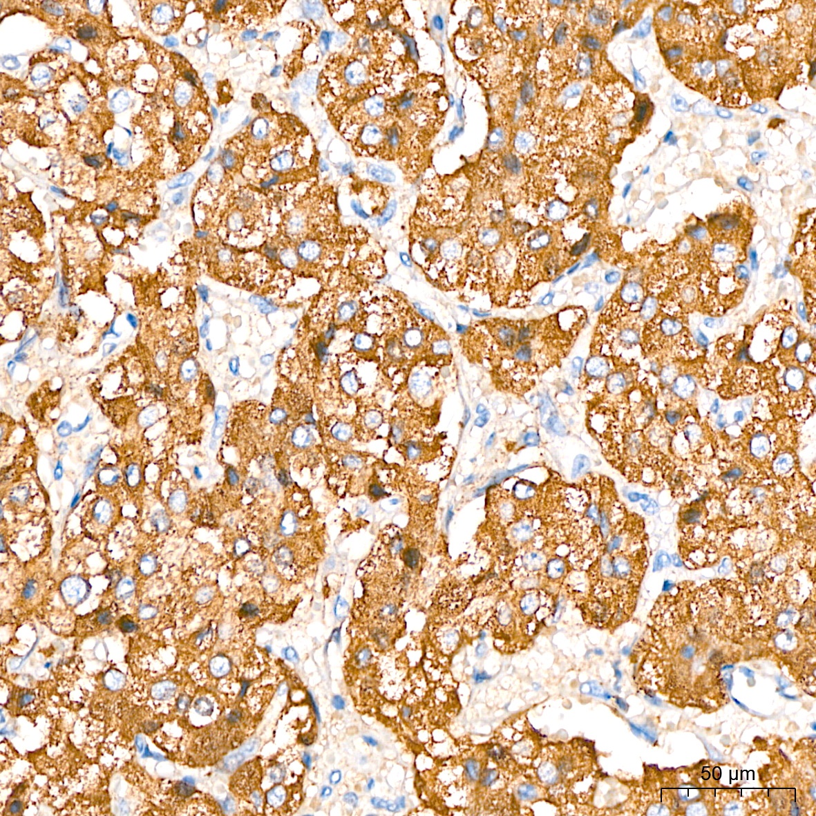
Immunohistochemistry analysis of paraffin-embedded Human pheochromocytoma tissue using Chromogranin A Rabbit mAb (CAB9576) at a dilution of 1:4000 (40x lens). High pressure antigen retrieval performed with 0.01M Citrate Buffer (pH 6.0) prior to IHC staining.
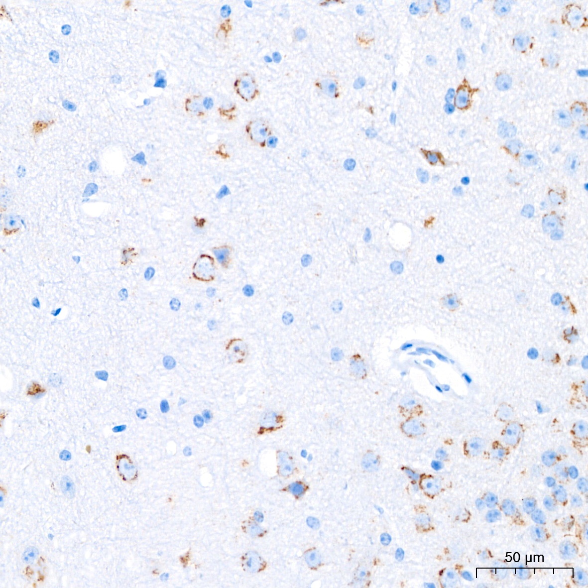
Immunohistochemistry analysis of paraffin-embedded Mouse brain tissue using Chromogranin A Rabbit mAb (CAB9576) at a dilution of 1:4000 (40x lens). High pressure antigen retrieval performed with 0.01M Citrate Buffer (pH 6.0) prior to IHC staining.
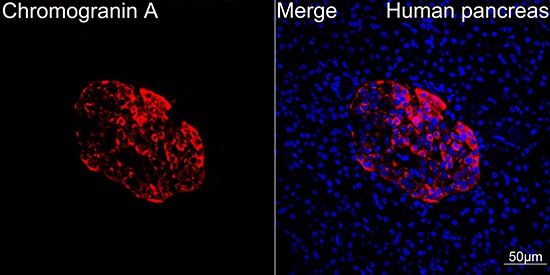
Confocal imaging of paraffin-embedded Human pancreas tissue using Chromogranin A Rabbit mAb (CAB9576, dilution 1:100) followed by a further incubation with Cy3 Goat Anti-Rabbit IgG (H+L) (CABS007, dilution 1:500) (Red). DAPI was used for nuclear staining (Blue). High pressure antigen retrieval performed with 0.01M Citrate Buffer (pH 6.0) prior to IF staining. Objective: 40x.
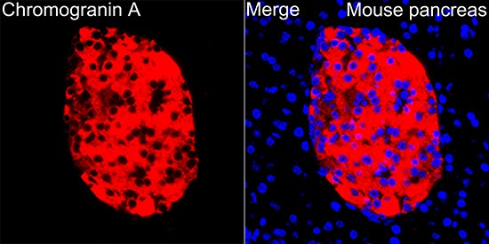
Confocal imaging of paraffin-embedded Mouse pancreas tissue using Chromogranin A Rabbit mAb (CAB9576, dilution 1:100) followed by a further incubation with Cy3 Goat Anti-Rabbit IgG (H+L) (CABS007, dilution 1:500) (Red). DAPI was used for nuclear staining (Blue). High pressure antigen retrieval performed with 0.01M Citrate Buffer (pH 6.0) prior to IF staining. Objective: 40x.
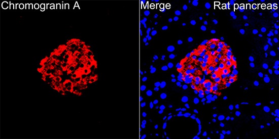
Confocal imaging of paraffin-embedded Rat pancreas tissue using Chromogranin A Rabbit mAb (CAB9576, dilution 1:100) followed by a further incubation with Cy3 Goat Anti-Rabbit IgG (H+L) (CABS007, dilution 1:500) (Red). DAPI was used for nuclear staining (Blue). High pressure antigen retrieval performed with 0.01M Citrate Buffer (pH 6.0) prior to IF staining. Objective: 40x.