The COPS3 Monoclonal Antibody (CAB19584) is a high-quality antibody developed for reliable detection and analysis of target proteins. This antibody, raised in rabbits, is highly specific and reactive with human samples, making it an ideal tool for studies in molecular biology and signal transduction pathways.COPS3, also known as COP9 signalosome subunit 3, plays a crucial role in the ubiquitin-proteasome system, regulating protein degradation and influencing various cellular processes. Its involvement in protein turnover and cell cycle control highlights its importance in cell function and homeostasis.
This antibody is validated for use in WB, IHC-P, ELISA applications and has demonstrated reactivity against Human, Mouse, Rat samples.
Product Name:
COPS3 Monoclonal Antibody
SKU:
CAB19584
Size:
20μL, 100μL
Reactivity:
Human, Mouse, Rat
Clone Number:
ARC2182
Conjugate:
Unconjugated
Immunogen:
Recombinant protein (or fragment).This information is considered to be commercially sensitive.
Cytoplasm, Cytosol, Nucleoplasm, Nucleus, Perinuclear Region Of Cytoplasm.
Calculated MW:
48kDa
Observed MW:
45kDa
The protein encoded by this gene possesses kinase activity that phosphorylates regulators involved in signal transduction. It phosphorylates I kappa-Balpha, p105, and c-Jun. It acts as a docking site for complex-mediated phosphorylation. The gene is located within the Smith-Magenis syndrome region on chromosome 17. Several transcript variants encoding different isoforms have been found for this gene.
Purification Method
Affinity purification
Gene ID
8533
Buffer Information
Store at -20℃. Avoid freeze / thaw cycles. Buffer: PBS containing 50% glycerol and 0.05% BSA, preserved with proclin300 or sodium azide, pH 7.3.
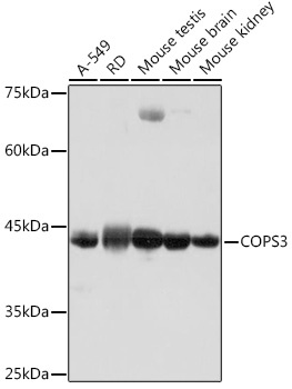
Western blot analysis of various lysates using COPS3 Rabbit mAb (CAB19584) at 1:1000 dilution. Secondary antibody: HRP-conjugated Goat anti-Rabbit IgG (H+L) (CABS014) at 1:10000 dilution. Lysates/proteins: 25μg per lane. Blocking buffer: 3% nonfat dry milk in TBST. Detection: ECL Basic Kit (AbGn00020). Exposure time: 1s.
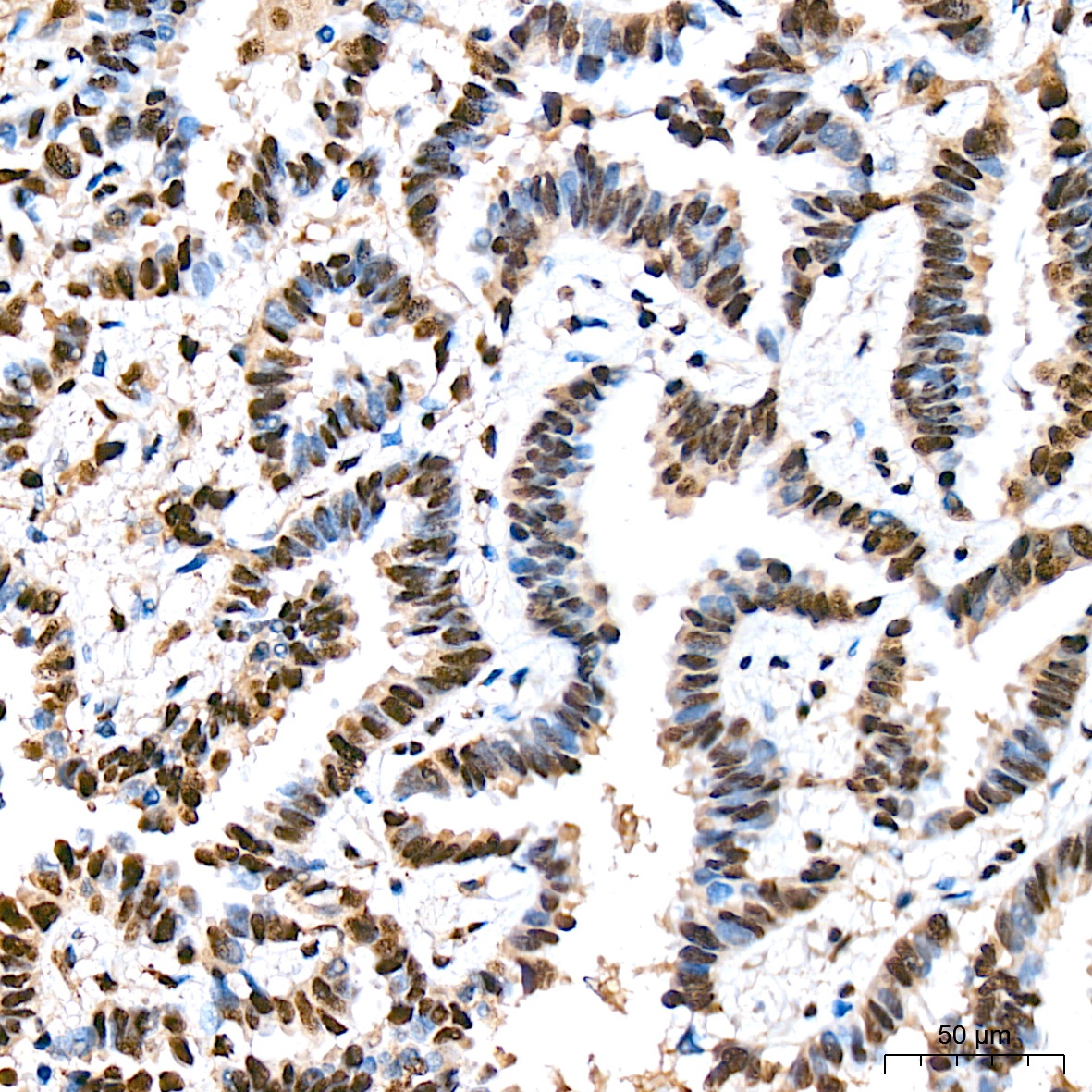
Immunohistochemistry analysis of paraffin-embedded Human lung cancer tissue using COPS3 Rabbit mAb (CAB19584) at a dilution of 1:200 (40x lens). High pressure antigen retrieval was performed with 0.01 M citrate buffer (pH 6.0) prior to IHC staining.
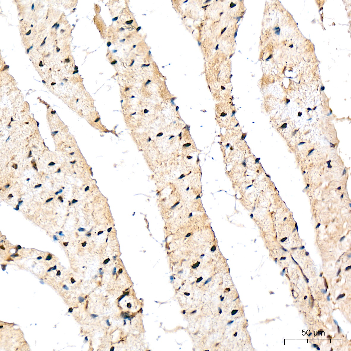
Immunohistochemistry analysis of paraffin-embedded Rat heart tissue using COPS3 Rabbit mAb (CAB19584) at a dilution of 1:200 (40x lens). High pressure antigen retrieval was performed with 0.01 M citrate buffer (pH 6.0) prior to IHC staining.