The ERp19 Monoclonal Antibody (CAB9386) is a high-quality antibody developed for reliable detection and analysis of target proteins. This polyclonal antibody, generated in rabbits, is suitable for detecting and analyzing ERP19 in human samples, making it a valuable tool for studies in cell biology, protein folding, and ER stress response.ERP19 is a key player in maintaining ER homeostasis by assisting in protein folding and quality control processes. Dysregulation of ERP19 expression or function has been linked to ER stress-related diseases such as neurodegenerative disorders, diabetes, and cardiovascular diseases.
This antibody is validated for use in WB, IHC-P, ELISA applications and has demonstrated reactivity against Human, Rat samples.
Product Name:
ERp19 Monoclonal Antibody
SKU:
CAB9386
Size:
20μL, 100μL
Reactivity:
Human, Rat
Clone Number:
ARC2718
Conjugate:
Unconjugated
Immunogen:
Synthetic peptide. This information is considered to be commercially sensitive.
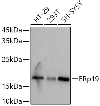
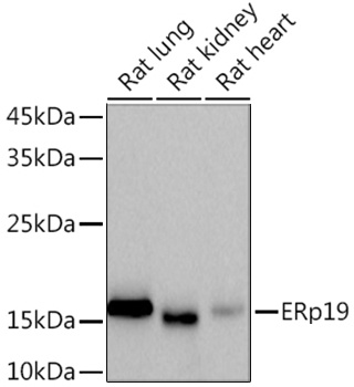
HT-29, 293T, SH-SY5Y, Rat lung, Rat kidney, Rat heart
Cellular Localization:
Endoplasmic Reticulum Lumen.
Calculated MW:
19kDa
Observed MW:
19kDa
This gene encodes a member of the thioredoxin superfamily. Members of this family are characterized by a conserved active motif called the thioredoxin fold that catalyzes disulfide bond formation and isomerization. This protein localizes to the endoplasmic reticulum and has a single atypical active motif. The encoded protein is mainly involved in catalyzing native disulfide bond formation and displays activity similar to protein-disulfide isomerases. This protein may play a role in defense against endoplasmic reticulum stress. Alternate splicing results in both coding and non-coding variants.
Purification Method
Affinity purification
Gene ID
51060
Buffer Information
Store at -20℃. Avoid freeze / thaw cycles. Buffer: PBS containing 50% glycerol and 0.05% BSA, preserved with proclin300 or sodium azide, pH 7.3.
Western blot analysis of various lysates using (CAB9386) at 1:1000 dilution. Secondary antibody: HRP-conjugated Goat anti-Rabbit IgG (H+L) (CABS014) at 1:10000 dilution. Lysates/proteins: 25μg per lane. Blocking buffer: 3% nonfat dry milk in TBST. Detection: ECL Basic Kit (AbGn00020). Exposure time: 3s.
Western blot analysis of various lysates using (CAB9386) at 1:1000 dilution. Secondary antibody: HRP-conjugated Goat anti-Rabbit IgG (H+L) (CABS014) at 1:10000 dilution. Lysates/proteins: 25μg per lane. Blocking buffer: 3% nonfat dry milk in TBST. Detection: ECL Basic Kit (AbGn00020). Exposure time: 90s.
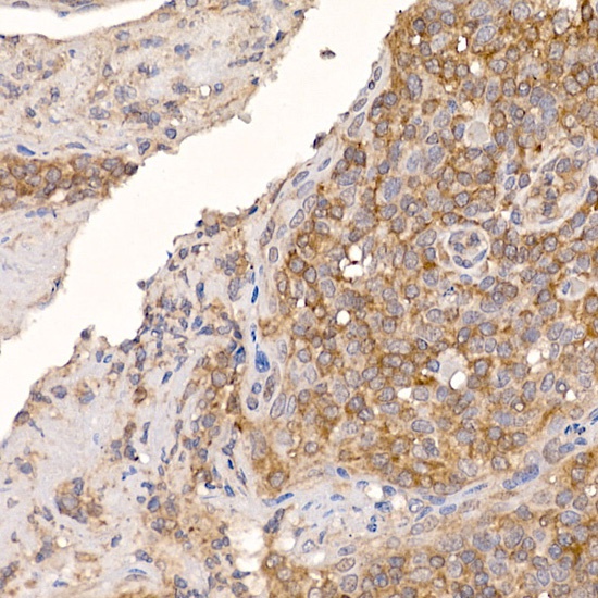
Immunohistochemistry analysis of paraffin-embedded Human tonsil using ERp19 Rabbit mAb (CAB9386) at dilution of 1:200 (40x lens). High pressure antigen retrieval performed with 0.01M Citrate buffer (pH 6.0) prior to IHC staining.
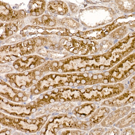
Immunohistochemistry analysis of paraffin-embedded Mouse kidney using ERp19 Rabbit mAb (CAB9386) at dilution of 1:200 (40x lens). High pressure antigen retrieval performed with 0.01M Citrate buffer (pH 6.0) prior to IHC staining.
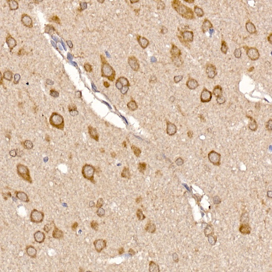
Immunohistochemistry analysis of paraffin-embedded Rat brain using ERp19 Rabbit mAb (CAB9386) at dilution of 1:200 (40x lens). High pressure antigen retrieval performed with 0.01M Citrate buffer (pH 6.0) prior to IHC staining.
. Perform high pressure antigen retrieval with 10 mM citrate buffer pH 6. 0 before commencing with IHC staining protocol.")
. Perform high pressure antigen retrieval with 10 mM citrate buffer pH 6. 0 before commencing with IHC staining protocol.")
. Perform high pressure antigen retrieval with 10 mM citrate buffer pH 6. 0 before commencing with IHC staining protocol.")
. Perform high pressure antigen retrieval with 10 mM citrate buffer pH 6. 0 before commencing with IHC staining protocol.")
. Perform high pressure antigen retrieval with 10 mM citrate buffer pH 6. 0 before commencing with IHC staining protocol.")
 at 1:10000 dilution. Lysates/proteins: 25ug per lane. Blocking buffer: 3% nonfat dry milk in TBST. Detection: ECL Basic Kit. Exposure time: 90s.")
 at 1:10000 dilution. Lysates/proteins: 25ug per lane. Blocking buffer: 3% nonfat dry milk in TBST. Detection: ECL Basic Kit. Exposure time: 3s.")
. Perform high pressure antigen retrieval with 10 mM citrate buffer pH 6. 0 before commencing with IHC staining protocol.")