The Ki67 Antibody (CAB11390) is a high-quality antibody developed for reliable detection and analysis of target proteins. This antibody, produced in rabbits, demonstrates high reactivity with human samples and has been validated for use in Western blot applications.The MKI67 Polyclonal Antibody binds specifically to the MKI67 protein, allowing for accurate detection and analysis in a variety of cell types. Its use is particularly beneficial in studies of cell proliferation, cell cycle regulation, and tumor growth, making it an essential tool for researchers in the fields of oncology, cell biology, and molecular biology.
This antibody is validated for use in IF/ICC, ELISA applications and has demonstrated reactivity against Human samples.
Product Name:
Ki67 Antibody
SKU:
CAB11390
Size:
20μL, 100μL
Reactivity:
Human
Conjugate:
Unconjugated
Immunogen:
Synthetic peptide. This information is considered to be commercially sensitive.
Recommended starting concentration is 1 μg/mL. Please optimize the concentration based on your specific assay requirements.
Synonyms:
KIA, MIB-, MIB-1, PPP1R105, Ki67
Positive Sample:
22Rv1, Raji
Cellular Localization:
Chromosome, Nucleus, Nucleolus.
Calculated MW:
359kDa
Observed MW:
359kDa
Enables protein C-terminus binding activity. Involved in regulation of chromosome segregation and regulation of mitotic nuclear division. Located in chromosome; nuclear body; and nucleolus. Colocalizes with condensed chromosome. Implicated in Crohn's disease; breast cancer; human immunodeficiency virus infectious disease; and pancreatic cancer. Biomarker of several diseases, including Barrett's esophagus; autoimmune disease of musculoskeletal system (multiple); endocrine gland cancer (multiple); gastrointestinal system cancer (multiple); and interstitial cystitis.
Purification Method
Affinity purification
Gene ID
4288
RRID
AB_2758539
Buffer Information
Store at -20℃. Avoid freeze / thaw cycles. Buffer: PBS containing 50% glycerol, preserved with proclin300 or sodium azide, pH 7.3.
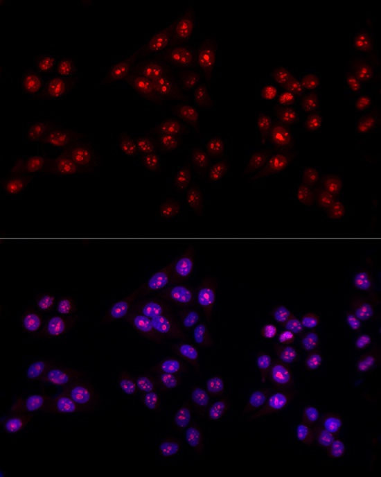
Immunofluorescence analysis of HeLa cells using Ki67 Rabbit pAb (CAB11390) at dilution of 1:100 (40x lens). Secondary antibody: Cy3-conjugated Goat anti-Rabbit IgG (H+L) (CABS007) at 1:500 dilution. Blue: DAPI for nuclear staining.


")

")


