The Histone H3.3 Antibody (CAB13824) is a high-quality antibody developed for reliable detection and analysis of target proteins. This antibody, raised in rabbits, is highly specific for human samples and has been validated for use in applications such as immunofluorescence and immunohistochemistry.H3F3A plays a crucial role in epigenetic regulation, DNA repair, and maintaining genomic stability. Dysregulation of H3F3A expression has been linked to various diseases, including cancer and neurological disorders.
This antibody is validated for use in WB, ELISA applications and has demonstrated reactivity against Human, Mouse, Rat, Other (Wide Range Predicted) samples.
Product Name:
Histone H3.3 Antibody
SKU:
CAB13824
Size:
20μL, 100μL
Reactivity:
Human, Mouse, Rat, Other (Wide Range Predicted)
Conjugate:
Unconjugated
Immunogen:
Recombinant protein (or fragment).This information is considered to be commercially sensitive.
Recommended starting concentration is 1 μg/mL. Please optimize the concentration based on your specific assay requirements.
Synonyms:
H3F3, H3-3B, H3.3A, H3F3A, BRYLIB1, Histone H3.3
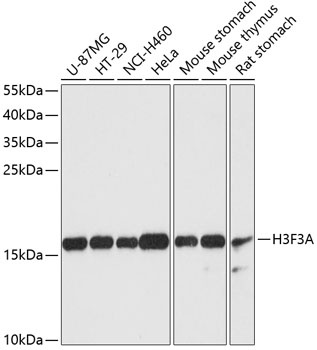
Positive Sample:
U-87MG, HT-29, NCI-H460, HeLa, Mouse stomach, Mouse thymus, Rat stomach
Cellular Localization:
Chromosome, Nucleus.
Calculated MW:
15kDa
Observed MW:
16kDa
Histones are basic nuclear proteins that are responsible for the nucleosome structure of the chromosomal fiber in eukaryotes. Two molecules of each of the four core histones (H2A, H2B, H3, and H4) form an octamer, around which approximately 146 bp of DNA is wrapped in repeating units, called nucleosomes. The linker histone, H1, interacts with linker DNA between nucleosomes and functions in the compaction of chromatin into higher order structures. This gene contains introns and its mRNA is polyadenylated, unlike most histone genes. The protein encoded is a replication-independent member of the histone H3 family.
Purification Method
Affinity purification
Gene ID
8290 8350
RRID
AB_2760678
Buffer Information
Store at -20℃. Avoid freeze / thaw cycles. Buffer: PBS with 0.01% thimerosal,50% glycerol,pH7.3.
Western blot analysis of various lysates using Histone H3.3 Rabbit pAb (CAB13824) at 1:3000 dilution. Secondary antibody: HRP-conjugated Goat anti-Rabbit IgG (H+L) (CABS014) at 1:10000 dilution. Lysates/proteins: 25μg per lane. Blocking buffer: 3% nonfat dry milk in TBST. Detection: ECL Basic Kit (AbGn00020). Exposure time: 90s.