The p53 DINP1 Monoclonal Antibody (CAB5952) is a high-quality antibody developed for reliable detection and analysis of target proteins. This polyclonal antibody, produced in rabbits, is validated for use in various applications, including Western blotting and immunofluorescence.The p53 protein, also known as the guardian of the genome, plays a central role in controlling cell cycle progression, DNA repair, and apoptosis in response to cellular stress. Mutations in p53 are commonly found in many human cancers, highlighting its importance in maintaining genomic stability and preventing malignant transformation.
This antibody is validated for use in WB, IHC-P, IF/ICC, ELISA applications and has demonstrated reactivity against Human, Mouse, Rat samples.
Product Name:
p53 DINP1 Monoclonal Antibody
SKU:
CAB5952
Size:
20μL, 100μL
Reactivity:
Human, Mouse, Rat
Clone Number:
ARC2102
Conjugate:
Unconjugated
Immunogen:
Synthetic peptide. This information is considered to be commercially sensitive.
Predicted to enable antioxidant activity. Involved in autophagic cell death; positive regulation of autophagy; and positive regulation of transcription, DNA-templated. Located in autophagosome; cytosol; and nucleus.
Purification Method
Affinity purification
Gene ID
94241
Buffer Information
Store at -20℃. Avoid freeze / thaw cycles. Buffer: PBS containing 50% glycerol and 0.05% BSA, preserved with proclin300 or sodium azide, pH 7.3.
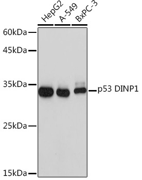
Western blot analysis of various lysates using p53 DINP1 Rabbit mAb (CAB5952) at 1:1000 dilution. Secondary antibody: HRP-conjugated Goat anti-Rabbit IgG (H+L) (CABS014) at 1:10000 dilution. Lysates/proteins: 25μg per lane. Blocking buffer: 3% nonfat dry milk in TBST. Detection: ECL Basic Kit (AbGn00020). Exposure time: 1s.
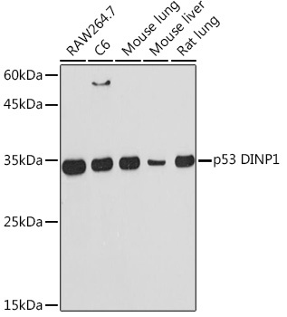
Western blot analysis of various lysates using p53 DINP1 Rabbit mAb (CAB5952) at 1:1000 dilution. Secondary antibody: HRP-conjugated Goat anti-Rabbit IgG (H+L) (CABS014) at 1:10000 dilution. Lysates/proteins: 25μg per lane. Blocking buffer: 3% nonfat dry milk in TBST. Detection: ECL Basic Kit (AbGn00020). Exposure time: 60s.
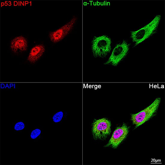
Confocal imaging of HeLa cells using p53 DINP1 Rabbit mAb (CAB5952, dilution 1:200) followed by a further incubation with Cy3 Goat Anti-Rabbit IgG (H+L) (CABS007,dilution 1:500) (Red). The cells were counterstained with α-Tubulin Mouse mAb (AC012, dilution 1:400) followed by incubation with ABflo® 488-conjugated Goat Anti-Mouse IgG (H+L) Ab (CABS076, dilution 1:500) (Green). DAPI was used for nuclear staining (Blue). Objective: 100x.