The p70 S6 Kinase 2 Monoclonal Antibody (CAB9100) is a high-quality antibody developed for reliable detection and analysis of target proteins. This antibody, produced in rabbits, exhibits strong reactivity with human samples and has been validated for use in Western blotting applications. By binding specifically to the p70 S6 kinase 2 protein, this antibody enables the detection and analysis of this critical signaling molecule in a variety of cell types, making it an excellent choice for studies in the fields of molecular biology and cancer research.p70 S6 kinase 2 plays a key role in the regulation of cell growth, cell proliferation, and protein synthesis in response to various stimuli.
This antibody is validated for use in WB, IHC-P, IF/ICC, ELISA applications and has demonstrated reactivity against Human, Mouse, Rat samples.
Product Name:
p70 S6 Kinase 2 Monoclonal Antibody
SKU:
CAB9100
Size:
20μL, 100μL
Reactivity:
Human, Mouse, Rat
Clone Number:
ARC1790
Conjugate:
Unconjugated
Immunogen:
Recombinant protein (or fragment).This information is considered to be commercially sensitive.
Sequence:
MAAV FDLD LETE EGSE GEGE PELS PADA CPLA ELRA AGLE PVGH YEEV ELTE TSVN VGPE RIGP HCFE LLRV LGKG GYGK VFQV RKVQ GTNL GKIY AMKV LRKA KIVR NAKD TAHT RAER
Tested Applications:
WBIHC-PIF/ICCELISA
Recommended Dilution:
WB
1:500 - 1:2000
IHC-P
1:50 - 1:200
IF/ICC
1:50 - 1:200
ELISA
Recommended starting concentration is 1 μg/mL. Please optimize the concentration based on your specific assay requirements.
This gene encodes a member of the RSK (ribosomal S6 kinase) family of serine/threonine kinases. This kinase contains a kinase catalytic domain and phosphorylates the S6 ribosomal protein and eukaryotic translation initiation factor 4B (eIF4B). Phosphorylation of S6 leads to an increase in protein synthesis and cell proliferation.
Purification Method
Affinity purification
Gene ID
6199
RRID
AB_2863656
Buffer Information
Store at -20℃. Avoid freeze / thaw cycles. Buffer: PBS containing 50% glycerol and 0.05% BSA, preserved with proclin300 or sodium azide, pH 7.3.
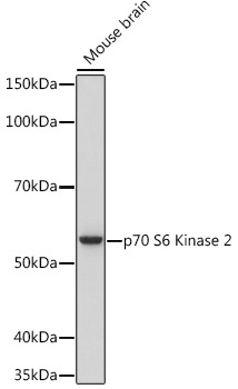
Western blot analysis of lysates from Mouse brain, using p70 S6 Kinase 2 Rabbit mAb (CAB9100) at 1:1000 dilution incubated overnight at 4℃. Secondary antibody: HRP-conjugated Goat anti-Rabbit IgG (H+L) (CABS014) at 1:10000 dilution. Lysates/proteins: 25 μg per lane. Blocking buffer: 3% nonfat dry milk in TBST. Detection: ECL Basic Kit (AbGn00020). Exposure time: 3 s.
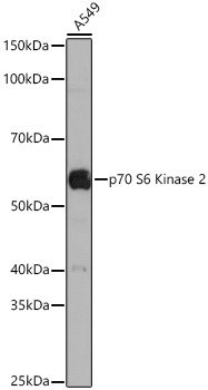
Western blot analysis of lysates from A549 cells using p70 S6 Kinase 2 Rabbit mAb (CAB9100) at 1:1000 dilution incubated overnight at 4℃. Secondary antibody: HRP-conjugated Goat anti-Rabbit IgG (H+L) (CABS014) at 1:10000 dilution. Lysates/proteins: 25 μg per lane. Blocking buffer: 3% nonfat dry milk in TBST. Detection: ECL Basic Kit (AbGn00020). Exposure time: 30 s.
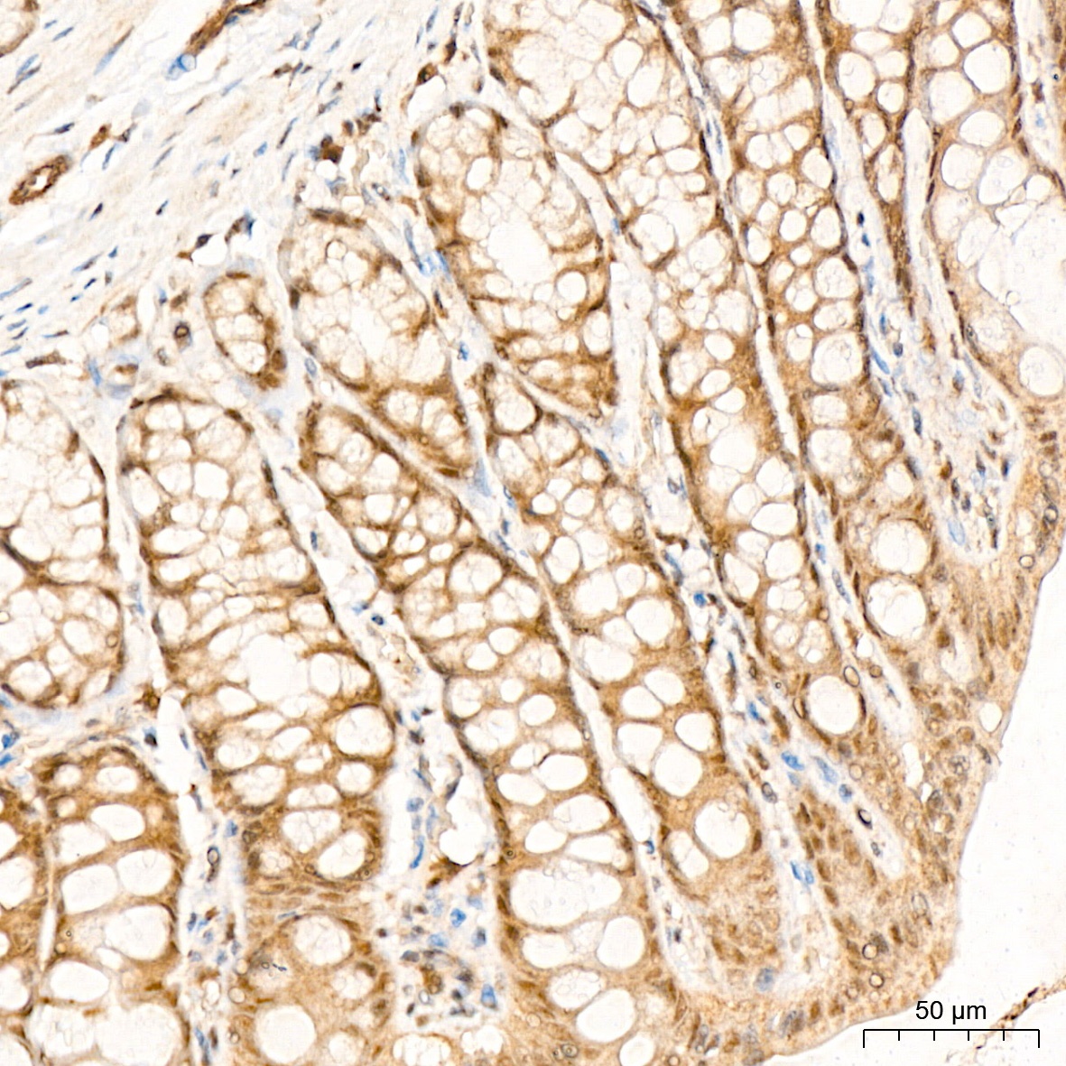
Immunohistochemistry analysis of paraffin-embedded Rat colon tissue using p70 S6 Kinase 2 Rabbit mAb (CAB9100) at a dilution of 1:200 (40x lens). High pressure antigen retrieval was performed with 0.01 M citrate buffer (pH 6.0) prior to IHC staining.
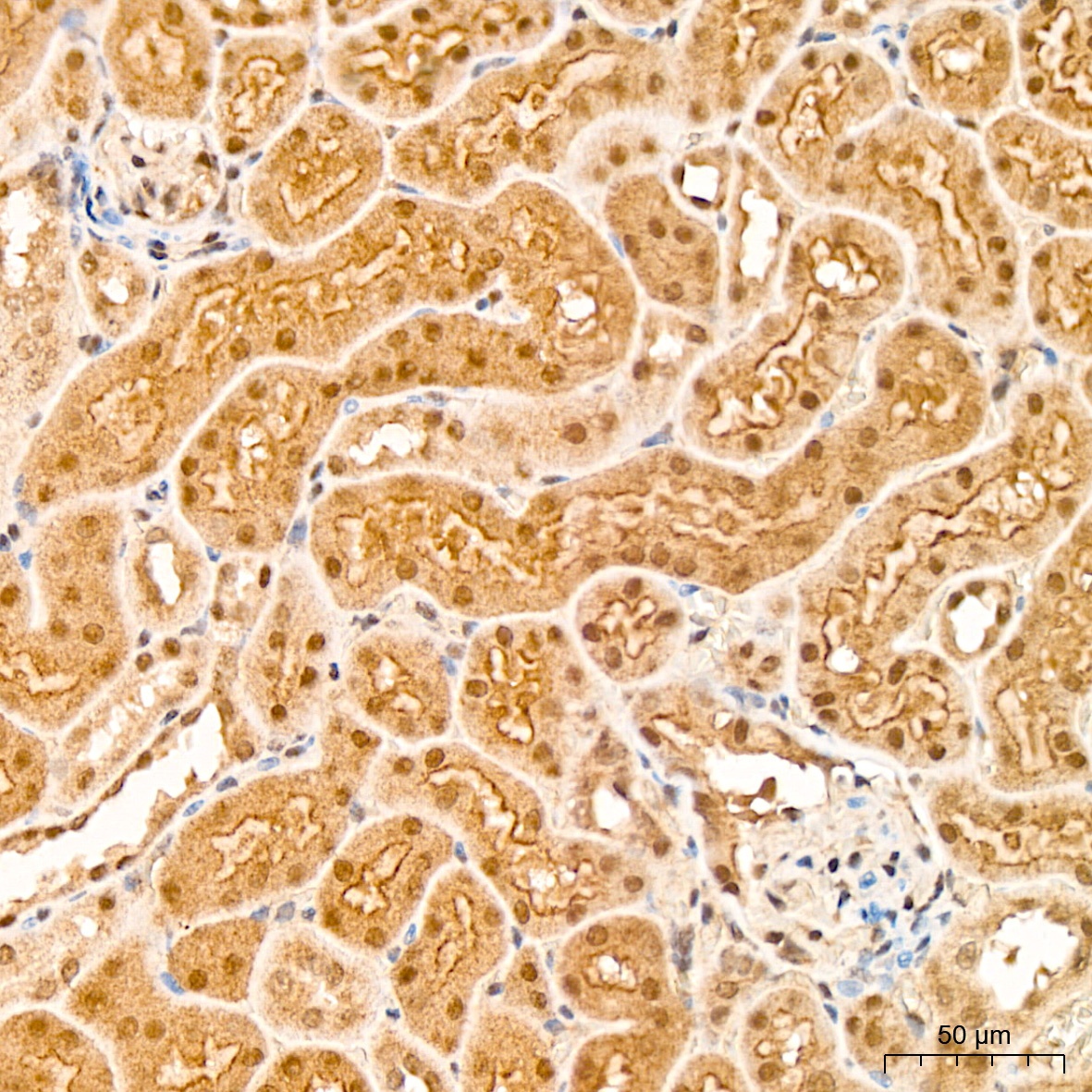
Immunohistochemistry analysis of paraffin-embedded Mouse kidney tissue using p70 S6 Kinase 2 Rabbit mAb (CAB9100) at a dilution of 1:200 (40x lens). High pressure antigen retrieval was performed with 0.01 M citrate buffer (pH 6.0) prior to IHC staining.
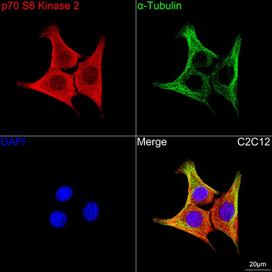
Confocal imaging of C2C12 cells using p70 S6 Kinase 2 Rabbit mAb (CAB9100, dilution 1:100) followed by a further incubation with Cy3 Goat Anti-Rabbit IgG (H+L) (CABS007, dilution 1:500) (Red). The cells were counterstained with α-Tubulin Mouse mAb (AC012, dilution 1:400) followed by incubation with ABflo® 488-conjugated Goat Anti-Mouse IgG (H+L) Ab (CABS076, dilution 1:500) (Green). DAPI was used for nuclear staining (Blue). Objective: 100x.
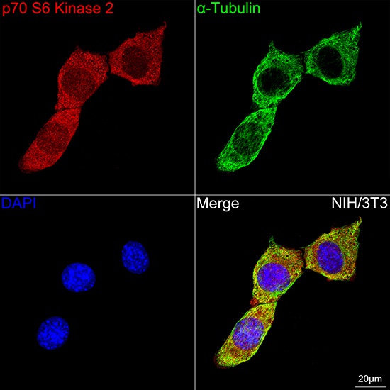
Confocal imaging of NIH/3T3 cells using p70 S6 Kinase 2 Rabbit mAb (CAB9100, dilution 1:100) followed by a further incubation with Cy3 Goat Anti-Rabbit IgG (H+L) (CABS007, dilution 1:500) (Red). The cells were counterstained with α-Tubulin Mouse mAb (AC012, dilution 1:400) followed by incubation with ABflo® 488-conjugated Goat Anti-Mouse IgG (H+L) Ab (CABS076, dilution 1:500) (Green). DAPI was used for nuclear staining (Blue). Objective: 100x.
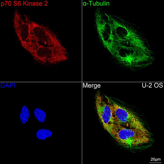
Confocal imaging of U-2 OS cells using p70 S6 Kinase 2 Rabbit mAb (CAB9100,dilution 1:100) followed by a further incubation with Cy3 Goat Anti-Rabbit IgG (H+L) (CABS007, dilution 1:500) (Red). The cells were counterstained with α-Tubulin Mouse mAb (AC012, dilution 1:400) followed by incubation with ABflo® 488-conjugated Goat Anti-Mouse IgG (H+L) Ab (CABS076, dilution 1:500) (Green). DAPI was used for nuclear staining (Blue). Objective: 100x.