The Pan Lactic acid-Lysine Antibody (CAB18831) is a high-quality antibody developed for reliable detection and analysis of target proteins. Histone lysine lactation (Kla) is a newly discovered histone modification that regulates gene expression in macrophages. In M1 macrophages, lactic acid is derived from incompletely oxidized glucose and then produces lactyl-CoA, which is transferred via acetyltransferase p300 to the lysine tail of the histone. This modification is abundant in gene promoter regions that lack acetylation and are associated with gene expression activation.
This antibody is validated for use in WB, ELISA applications and has demonstrated reactivity against Human, Mouse, Rat, Other (Wide Range) samples.
Product Name:
Pan Lactic acid-Lysine Antibody
SKU:
CAB18831
Size:
100μL, 20μL
Reactivity:
Human, Mouse, Rat, Other (Wide Range)
Immunogen:
Synthetic peptide. This information is considered to be commercially sensitive.
Tested Applications:
WBELISA
Recommended Dilution:
WB
1:500 - 1:1000
ELISA
Recommended starting concentration is 1 μg/mL. Please optimize the concentration based on your specific assay requirements.
Positive Sample:
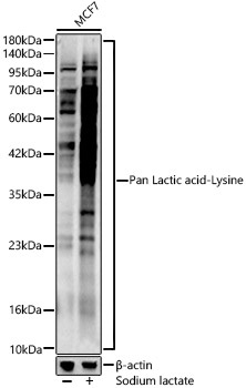
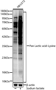
MCF7 treated with Sodium lactate, NIH/3T3 treated with Sodium lactate, C6 treated with Sodium lactate
Histone lysine lactation (Kla) is a newly discovered histone modification that regulates gene expression in macrophages. In M1 macrophages, lactic acid is derived from incompletely oxidized glucose and then produces lactyl-CoA, which is transferred via acetyltransferase p300 to the lysine tail of the histone. This modification is abundant in gene promoter regions that lack acetylation and are associated with gene expression activation.
Purification Method
Affinity purification
RRID
AB_2862466
Buffer Information
Store at -20℃. Avoid freeze / thaw cycles. Buffer: PBS with 0.09% sodium azide,50% glycerol,pH7.3.
Western blot analysis of lysates from MCF7 cells, using Pan Lactic acid-Lysine Rabbit pAb (CAB18831) at 1:400 dilution. MCF7 cells were treated with Sodium lactate(100mM) for 24h. Secondary antibody: HRP-conjugated Goat anti-Rabbit IgG (H+L) (AS014) at 1:10000 dilution. Lysates/proteins: 25μg per lane. Blocking buffer: 3% nonfat dry milk in TBST. Detection: ECL Enhanced Kit (AbGn00021). Exposure time: 60s.
Western blot analysis of lysates from NIH/3T3 cells, using Pan Lactic acid-Lysine Rabbit pAb (CAB18831) at 1:400 dilution. NIH/3T3 cells were treated with Sodium lactate(100mM) for 24h. Secondary antibody: HRP-conjugated Goat anti-Rabbit IgG (H+L) (AS014) at 1:10000 dilution. Lysates/proteins: 25μg per lane. Blocking buffer: 3% nonfat dry milk in TBST. Detection: ECL Enhanced Kit (AbGn00021). Exposure time: 60s.