The PDLIM4 Antibody (CAB20463) is a high-quality antibody developed for reliable detection and analysis of target proteins. This antibody, produced in rabbits, demonstrates high reactivity with human samples and has been validated for use in Western blot experiments. By specifically binding to the PDLIM4 protein, this antibody facilitates the detection and analysis of PDLIM4 in various cell types, making it ideal for investigations in cell biology and cancer research.PDLIM4, also known as the PDZ and LIM domain protein 4, plays a crucial role in regulating cell growth, differentiation, and migration.
This antibody is validated for use in WB, IF/ICC, ELISA applications and has demonstrated reactivity against Human, Mouse, Rat samples.
Product Name:
PDLIM4 Antibody
SKU:
CAB20463
Size:
20μL, 100μL
Reactivity:
Human, Mouse, Rat
Conjugate:
Unconjugated
Immunogen:
Recombinant protein (or fragment).This information is considered to be commercially sensitive.
Recommended starting concentration is 1 μg/mL. Please optimize the concentration based on your specific assay requirements.
Synonyms:
RIL, PDLIM4
Positive Sample:
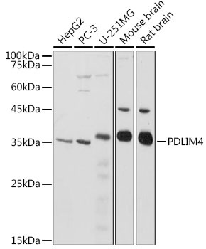
HepG2, PC-3, U-251MG, Mouse brain, Rat brain
Cellular Localization:
Cytoplasm, Cytoskeleton, Dendritic Spine, Early Endosome Lumen, Nucleus, Perinuclear Region Of Cytoplasm, Postsynaptic Membrane, Recycling Endosome Lumen, Z Disc.
Calculated MW:
35kDa
Observed MW:
35kDa
Enables alpha-actinin binding activity; protein homodimerization activity; and protein phosphatase binding activity. Involved in actin cytoskeleton reorganization. Located in several cellular components, including lamellipodium; perinuclear region of cytoplasm; and stress fiber. Part of filamentous actin. Implicated in osteoporosis.
Purification Method
Affinity purification
Gene ID
8572
Buffer Information
Store at -20℃. Avoid freeze / thaw cycles. Buffer: PBS containing 50% glycerol, preserved with proclin300 or sodium azide, pH 7.3.
Western blot analysis of various lysates using PDLIM4 Rabbit pAb (CAB20463) at 1:1000 dilution. Secondary antibody: HRP-conjugated Goat anti-Rabbit IgG (H+L) (CABS014) at 1:10000 dilution. Lysates/proteins: 25μg per lane. Blocking buffer: 3% nonfat dry milk in TBST. Detection: ECL Basic Kit (AbGn00020). Exposure time: 3s.
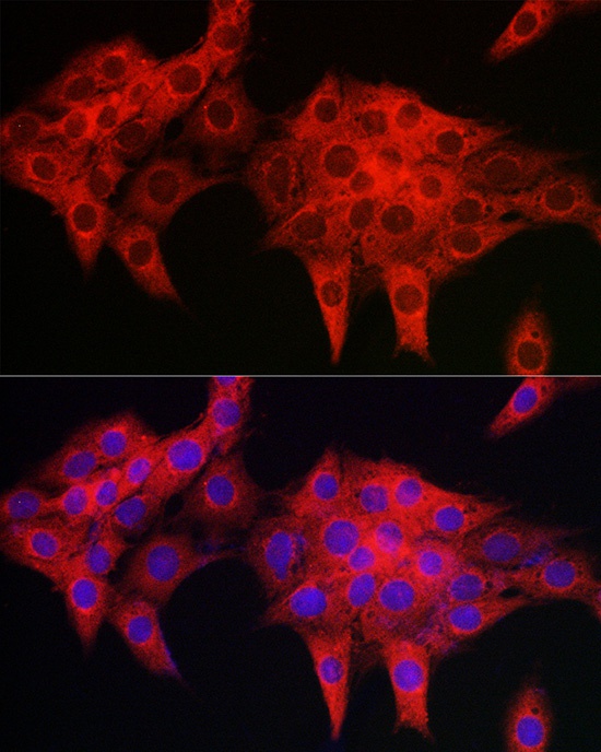
Immunofluorescence analysis of PC-12 cells using PDLIM4 Rabbit pAb (CAB20463) at dilution of 1:50 (40x lens). Secondary antibody: Cy3-conjugated Goat anti-Rabbit IgG (H+L) (CABS007) at 1:500 dilution. Blue: DAPI for nuclear staining.