The Phospholipid Scramblase 1 (PLSCR1) Monoclonal Antibody (CAB3430) is a high-quality antibody developed for reliable detection and analysis of target proteins. This polyclonal antibody, produced in rabbits, demonstrates high specificity and sensitivity in detecting PLSCR1 protein in various experimental settings, including Western blot analysis.PLSCR1 is a phospholipid scramblase enzyme that plays essential roles in lipid metabolism, cell signaling, and membrane dynamics. It is involved in various cellular processes, including cell proliferation, apoptosis, and inflammatory responses.
This antibody is validated for use in WB, IHC-P, IF/ICC, ELISA applications and has demonstrated reactivity against Human, Mouse, Rat samples.
Recommended starting concentration is 1 μg/mL. Please optimize the concentration based on your specific assay requirements.
Synonyms:
MMTRA1B, Phospholipid Scramblase 1 (PLSCR1)
Positive Sample:
HT-29, Mouse lung
Cellular Localization:
Cell Membrane, Nucleus, Cytoplasm, Cytoplasm, Perinuclear Region .
Calculated MW:
35kDa
Observed MW:
35kDa
This gene encodes a phospholipid scramblase family member. The encoded protein is involved in disruption of the asymmetrical distribution of phospholipids between the inner and outer leaflets of the plasma membrane, resulting in externalization of phosphatidylserine. This cell membrane disruption plays an important role in the blood coagulation cascade as well as macrophage clearing of apoptotic cells. The encoded protein has additionally been implicated in gene regulation and interferon-induced antiviral responses.
Purification Method
Affinity purification
Gene ID
5359
RRID
AB_2863056
Buffer Information
Store at -20℃. Avoid freeze / thaw cycles. Buffer: PBS containing 50% glycerol and 0.05% BSA, preserved with proclin300 or sodium azide, pH 7.3.
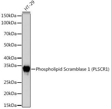
Western blot analysis of lysates from HT-29 cells, using Phospholipid Phospholipid Scramblase 1 (PLSCR1) (PLSCR1) Rabbit mAb (CAB3430) at 1:1000 dilution. Secondary antibody: HRP-conjugated Goat anti-Rabbit IgG (H+L) (CABS014) at 1:10000 dilution. Lysates/proteins: 25μg per lane. Blocking buffer: 3% nonfat dry milk in TBST. Detection: ECL Basic Kit (AbGn00020). Exposure time: 3s.
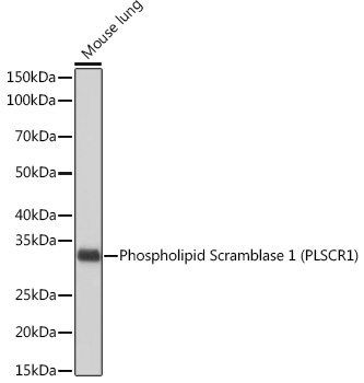
Western blot analysis of lysates from Mouse lung, using Phospholipid Phospholipid Scramblase 1 (PLSCR1) (PLSCR1) Rabbit mAb (CAB3430) at 1:1000 dilution. Secondary antibody: HRP-conjugated Goat anti-Rabbit IgG (H+L) (CABS014) at 1:10000 dilution. Lysates/proteins: 25μg per lane. Blocking buffer: 3% nonfat dry milk in TBST. Detection: ECL Basic Kit (AbGn00020). Exposure time: 10s.
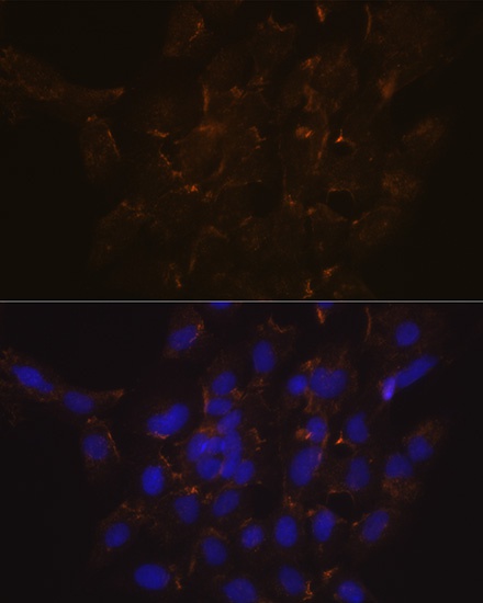
Immunofluorescence analysis of C6 cells using Phospholipid Phospholipid Scramblase 1 (PLSCR1) (PLSCR1) Rabbit mAb (CAB3430) at dilution of 1:100 (40x lens). Secondary antibody: Cy3-conjugated Goat anti-Rabbit IgG (H+L) (CABS007) at 1:500 dilution. Blue: DAPI for nuclear staining.


 ELISA Kit (HUEB1946)")
 ELISA Kit (RTEB1203)")
 ELISA Kit (MOEB1687)")
")