The PIK3R4/VPS15 Monoclonal Antibody (CAB5922) is a high-quality antibody developed for reliable detection and analysis of target proteins. This antibody, produced in rabbits, is highly specific for human samples and has been validated for use in Western blot applications.The PIK3R4/VPS15 protein is a key component of the class III phosphatidylinositol 3-kinase (PI3K) complex, which is involved in the regulation of autophagy, a process that helps cells maintain homeostasis by degrading and recycling damaged organelles and proteins.
This antibody is validated for use in WB, IF/ICC, ELISA applications and has demonstrated reactivity against Human, Mouse samples.
Product Name:
PIK3R4/VPS15 Monoclonal Antibody
SKU:
CAB5922
Size:
20μL, 100μL
Reactivity:
Human, Mouse
Clone Number:
ARC2090
Conjugate:
Unconjugated
Immunogen:
Synthetic peptide. This information is considered to be commercially sensitive.
Recommended starting concentration is 1 μg/mL. Please optimize the concentration based on your specific assay requirements.
Synonyms:
p150, VPS15, PIK3R4/VPS15
Positive Sample:
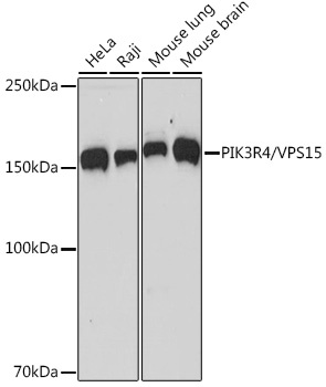
HeLa, Raji, Mouse lung, Mouse brain
Cellular Localization:
Autophagosome, Axoneme, Cytosol, Late Endosome, Microtubule Cytoskeleton.
Calculated MW:
153kDa
Observed MW:
153kDa
Predicted to enable protein serine/threonine kinase activity. Involved in positive regulation of phosphatidylinositol 3-kinase activity; receptor catabolic process; and regulation of cytokinesis. Located in late endosome and microtubule cytoskeleton.
Purification Method
Affinity purification
Gene ID
30849
Buffer Information
Store at -20℃. Avoid freeze / thaw cycles. Buffer: PBS containing 50% glycerol and 0.05% BSA, preserved with proclin300 or sodium azide, pH 7.3.
Western blot analysis of various lysates using PIK3R4/VPS15 Rabbit mAb (CAB5922) at 1:1000 dilution. Secondary antibody: HRP-conjugated Goat anti-Rabbit IgG (H+L) (CABS014) at 1:10000 dilution. Lysates/proteins: 25μg per lane. Blocking buffer: 3% nonfat dry milk in TBST. Detection: ECL Basic Kit (AbGn00020). Exposure time: 60s.
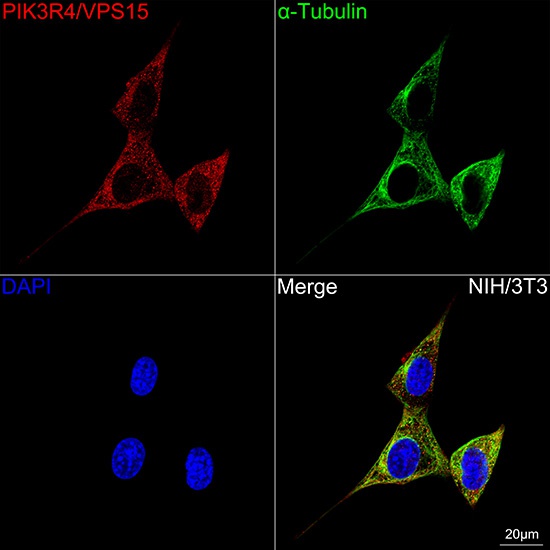
Confocal imaging of NIH/3T3 cells using PIK3R4/VPS15 Rabbit mAb (CAB5922, dilution 1:100) followed by a further incubation with Cy3 Goat Anti-Rabbit IgG (H+L) (CABS007, dilution 1:500) (Red). The cells were counterstained with α-Tubulin Mouse mAb (AC012, dilution 1:400) followed by incubation with ABflo® 488-conjugated Goat Anti-Mouse IgG (H+L) Ab (CABS076, dilution 1:500) (Green). DAPI was used for nuclear staining (Blue). Objective: 100x.