The Rev-Erbalpha/NR1D1 Monoclonal Antibody (CAB20452) is a high-quality antibody developed for reliable detection and analysis of target proteins. This antibody, generated in rabbits, is highly specific to human samples and has been validated for use in Western blot and immunohistochemistry applications.REV-ERBα, a member of the nuclear receptor superfamily, plays a crucial role in the control of physiological processes such as metabolism, inflammation, and the sleep-wake cycle. Dysregulation of REV-ERBα has been linked to various metabolic disorders, making it a promising target for therapeutic interventions.
This antibody is validated for use in WB, IF/ICC, ELISA, IF-P applications and has demonstrated reactivity against Human, Mouse, Rat samples.
Product Name:
Rev-Erbalpha/NR1D1 Monoclonal Antibody
SKU:
CAB20452
Size:
20μL, 100μL
Reactivity:
Human, Mouse, Rat
Clone Number:
ARC50483
Conjugate:
Unconjugated
Immunogen:
Recombinant protein (or fragment).This information is considered to be commercially sensitive.
This gene encodes a transcription factor that is a member of the nuclear receptor subfamily 1. The encoded protein is a ligand-sensitive transcription factor that negatively regulates the expression of core clock proteins. In particular this protein represses the circadian clock transcription factor aryl hydrocarbon receptor nuclear translocator-like protein 1 (ARNTL). This protein may also be involved in regulating genes that function in metabolic, inflammatory and cardiovascular processes.
Purification Method
Affinity purification
Gene ID
9572
Buffer Information
Store at -20℃. Avoid freeze / thaw cycles. Buffer: PBS containing 50% glycerol and 0.05% BSA, preserved with proclin300 or sodium azide, pH 7.3.
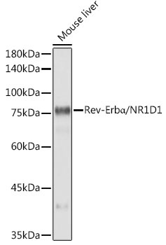
Western blot analysis of lysates from Mouse liver, using Rev-Erbα/NR1D1 Rabbit mAb (CAB20452) at1:1000 dilution. Secondary antibody: HRP-conjugated Goat anti-Rabbit IgG (H+L) (CABS014) at1:10000 dilution. Lysates/proteins: 25μg per lane. Blocking buffer: 3% nonfat dry milk in TBST. Detection: ECL Basic Kit (AbGn00020). Exposure time: 10s.
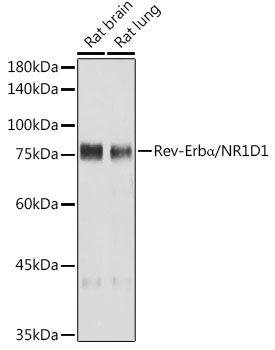
Western blot analysis of various lysates using Rev-Erbα/NR1D1 Rabbit mAb (CAB20452) at1:1000 dilution. Secondary antibody: HRP-conjugated Goat anti-Rabbit IgG (H+L) (CABS014) at1:10000 dilution. Lysates/proteins: 25μg per lane. Blocking buffer: 3% nonfat dry milk in TBST. Detection: ECL Basic Kit (AbGn00020). Exposure time: 30s.
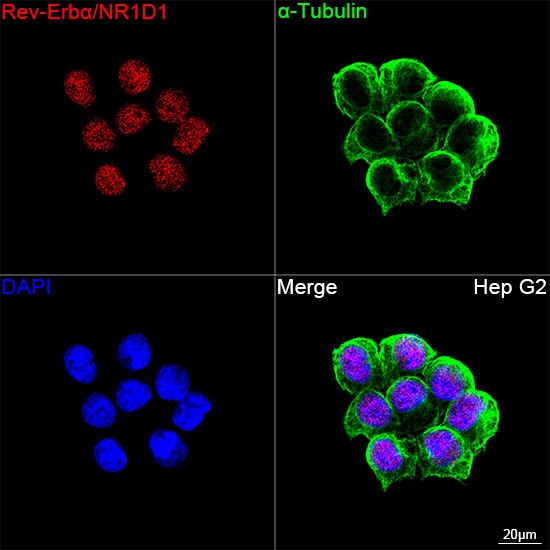
Confocal imaging of Hep G2 cells using Rev-Erbα/NR1D1 Rabbit mAb (CAB20452, dilution 1:200) followed by a further incubation with Cy3 Goat Anti-Rabbit IgG (H+L) (CABS007, dilution 1:500) (Red). The cells were counterstained with α-Tubulin Mouse mAb (AC012, dilution 1:400) followed by incubation with ABflo® 488-conjugated Goat Anti-Mouse IgG (H+L) Ab (CABS076, dilution 1:500) (Green). DAPI was used for nuclear staining (Blue). Objective: 100x.
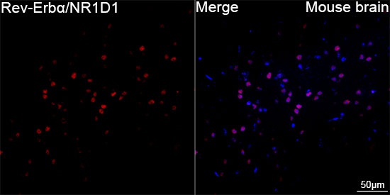
Confocal imaging of paraffin-embedded Mouse brain tissue using Rev-Erbα/NR1D1 Rabbit mAb (CAB20452, dilution 1:200) followed by a further incubation with Cy3 Goat Anti-Rabbit IgG (H+L) (CABS007, dilution 1:500) (Red). DAPI was used for nuclear staining (Blue). High pressure antigen retrieval performed with 0.01M Citrate Buffer (pH 6.0) prior to IF staining. Objective: 40x.