The STK39 Monoclonal Antibody (CAB2275) is a high-quality antibody developed for reliable detection and analysis of target proteins. This antibody, produced in rabbits, is highly specific to human samples and has been validated for use in Western blot applications. By targeting the STK39 protein, this antibody enables precise detection and analysis in a wide range of cell types, making it an essential component for studies in molecular biology and disease research.STK39, also known as STE20/SPS1-related proline/alanine-rich kinase, is critically involved in regulating ion channels and maintaining cellular homeostasis.
This antibody is validated for use in WB, IHC-P, IF/ICC, ELISA, IF-P applications and has demonstrated reactivity against Human, Mouse, Rat samples.
Product Name:
STK39 Monoclonal Antibody
SKU:
CAB2275
Size:
20μL, 100μL
Reactivity:
Human, Mouse, Rat
Clone Number:
ARC1896
Conjugate:
Unconjugated
Immunogen:
Recombinant protein (or fragment).This information is considered to be commercially sensitive.
Recommended starting concentration is 1 μg/mL. Please optimize the concentration based on your specific assay requirements.
Synonyms:
DCHT, PASK, SPAK, Ste 20 related kinase, STK39
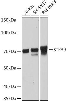
Positive Sample:
Jurkat, SH-SY5Y, Rat testis
Cellular Localization:
Cytoplasm, Nucleus.
Calculated MW:
59kDa
Observed MW:
68kDa
This gene encodes a serine/threonine kinase that is thought to function in the cellular stress response pathway. The kinase is activated in response to hypotonic stress, leading to phosphorylation of several cation-chloride-coupled cotransporters. The catalytically active kinase specifically activates the p38 MAP kinase pathway, and its interaction with p38 decreases upon cellular stress, suggesting that this kinase may serve as an intermediate in the response to cellular stress.
Purification Method
Affinity purification
Gene ID
27347
RRID
AB_2862985
Buffer Information
Store at -20℃. Avoid freeze / thaw cycles. Buffer: PBS containing 50% glycerol and 0.05% BSA, preserved with proclin300 or sodium azide, pH 7.3.
Western blot analysis of various lysates using STK39 Rabbit mAb (CAB2275) at 1:1000 dilution. Secondary antibody: HRP-conjugated Goat anti-Rabbit IgG (H+L) (CABS014) at 1:10000 dilution. Lysates/proteins: 25μg per lane. Blocking buffer: 3% nonfat dry milk in TBST. Detection: ECL Basic Kit (AbGn00020). Exposure time: 10s.
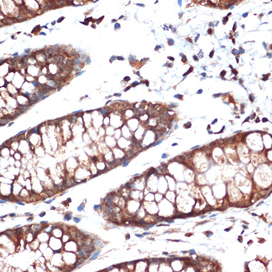
Immunohistochemistry analysis of paraffin-embedded Human colon using STK39 Rabbit mAb (CAB2275) at dilution of 1:100 (40x lens). Microwave antigen retrieval performed with 0.01M Tris/EDTA Buffer (pH 9.0) prior to IHC staining.
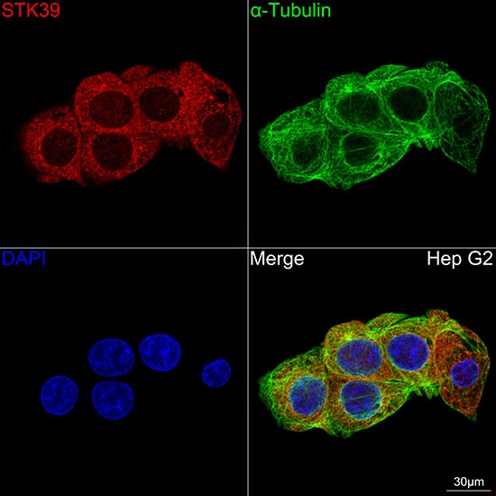
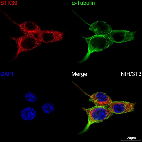
Confocal imaging of Hep G2 cells using STK39 Rabbit mAb (CAB2275,at dilution of 1:100) (Red). The cells were counterstained with α-Tubulin Mouse mAb (AC012,dilution 1:400) (Green). DAPI was used for nuclear staining (blue). DAPI was used for nuclear staining (blue). Objective: 100x.
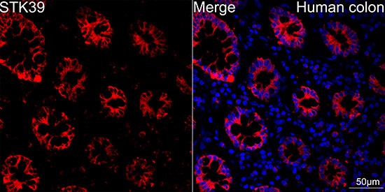
Confocal imaging of human colon using STK39 Rabbit mAb (CAB2275,at dilution of 1:100) (Red). Objective: 40x. Perform high pressure antigen retrieval with 10 mM citrate buffer pH 6.0 before commencing with IF staining protocol.
Western blot analysis of various lysates using STK39 Rabbit mAb (CAB2275) at 1:1000 dilution. Secondary antibody: HRP-conjugated Goat anti-Rabbit IgG (H+L) (CABS014) at 1:10000 dilution. Lysates/proteins: 25μg per lane. Blocking buffer: 3% nonfat dry milk in TBST. Detection: ECL Basic Kit (AbGn00020). Exposure time: 10s.