The SUN2 Monoclonal Antibody (CAB19782) is a high-quality antibody developed for reliable detection and analysis of target proteins. The antibody is raised in rabbits and validated for use in various applications, including Western blot and immunofluorescence.SUN2 is a key component of the linker of nucleoskeleton and cytoskeleton (LINC) complex, which plays important roles in nuclear migration, chromatin organization, and mechanotransduction. The Anti-SUN2 Antibody binds specifically to SUN2, allowing for the visualization and analysis of its expression and localization in different cell types.
This antibody is validated for use in WB, IHC-P, ELISA applications and has demonstrated reactivity against Human, Mouse, Rat samples.
Product Name:
SUN2 Monoclonal Antibody
SKU:
CAB19782
Size:
20μL, 100μL
Reactivity:
Human, Mouse, Rat
Clone Number:
ARC2311
Conjugate:
Unconjugated
Immunogen:
Synthetic peptide. This information is considered to be commercially sensitive.
Recommended starting concentration is 1 μg/mL. Please optimize the concentration based on your specific assay requirements.
Synonyms:
UNC84B, rab5IP, SUN2
Positive Sample:
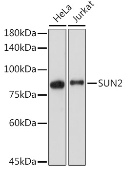
HeLa, Jurkat
Cellular Localization:
Nuclear Envelope, Nuclear Membrane.
Calculated MW:
80kDa
Observed MW:
80kDa
SUN1 (MIM 607723) and SUN2 are inner nuclear membrane (INM) proteins that play a major role in nuclear-cytoplasmic connection by formation of a 'bridge' across the nuclear envelope, known as the LINC complex, via interaction with the conserved luminal KASH domain of nesprins (e.g., SYNE1; MIM 608441) located in the outer nuclear membrane (ONM). The LINC complex provides a direct connection between the nuclear lamina and the cytoskeleton, which contributes to nuclear positioning and cellular rigidity (summary by Haque et al., 2010 [PubMed 19933576]).
Purification Method
Affinity purification
Gene ID
25777
Buffer Information
Store at -20℃. Avoid freeze / thaw cycles. Buffer: PBS containing 50% glycerol and 0.05% BSA, preserved with proclin300 or sodium azide, pH 7.3.
Western blot analysis of various lysates using SUN2 Rabbit mAb (CAB19782) at 1:2000 dilution. Secondary antibody: HRP-conjugated Goat anti-Rabbit IgG (H+L) (CABS014) at 1:10000 dilution. Lysates/proteins: 25μg per lane. Blocking buffer: 3% nonfat dry milk in TBST. Detection: ECL Enhanced Kit (AbGn00021). Exposure time: 60s.
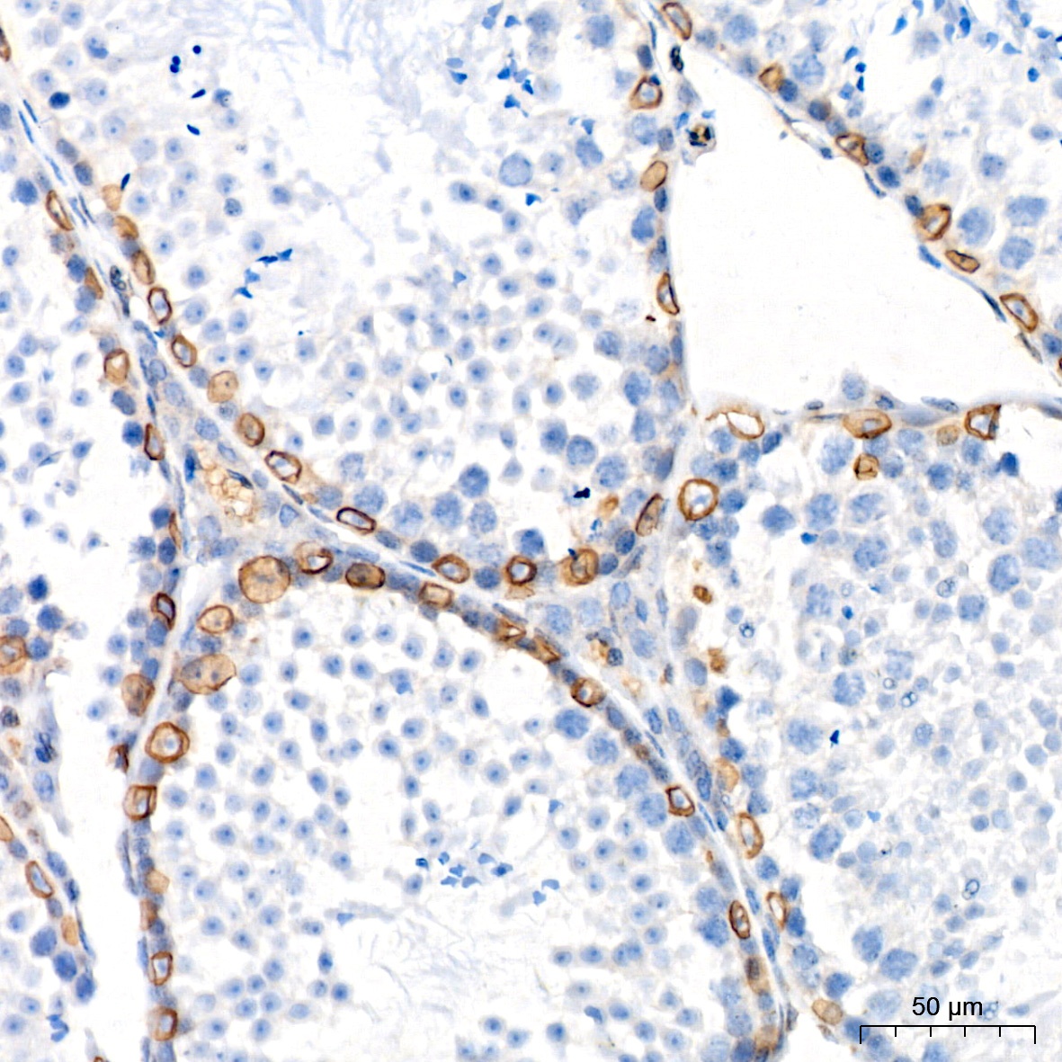
Immunohistochemistry analysis of paraffin-embedded Mouse testis tissue using SUN2 Rabbit mAb (CAB19782) at a dilution of 1:200 (40x lens). High pressure antigen retrieval performed with 0.01M Citrate buffer (pH 6.0) prior to IHC staining.
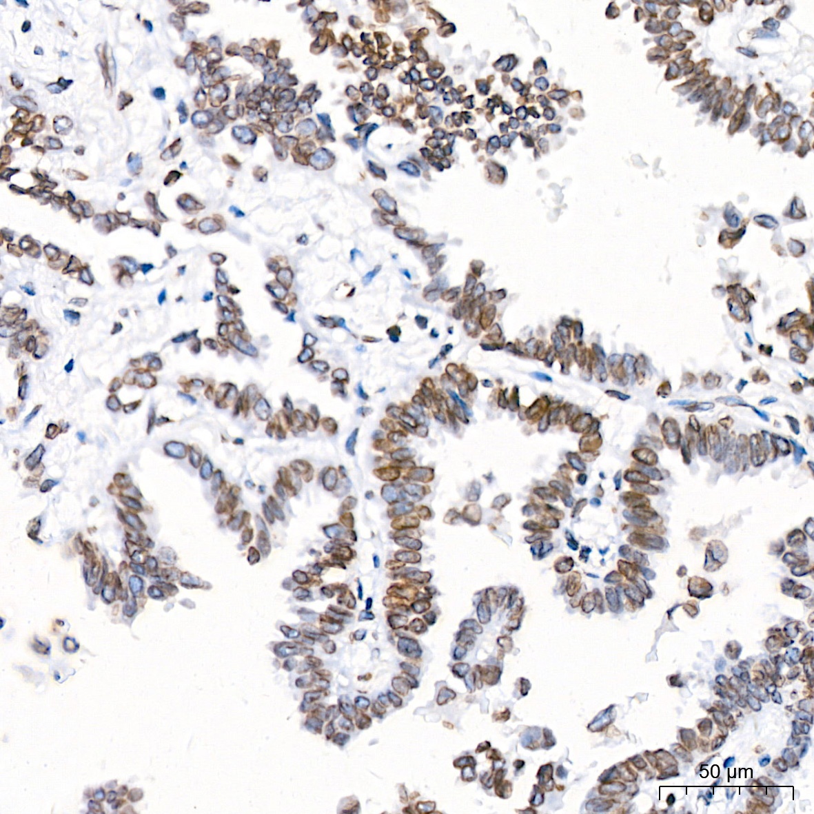
Immunohistochemistry analysis of paraffin-embedded Human lung adenocarcinoma tissue using SUN2 Rabbit mAb (CAB19782) at a dilution of 1:200 (40x lens). High pressure antigen retrieval performed with 0.01M Citrate buffer (pH 6.0) prior to IHC staining.
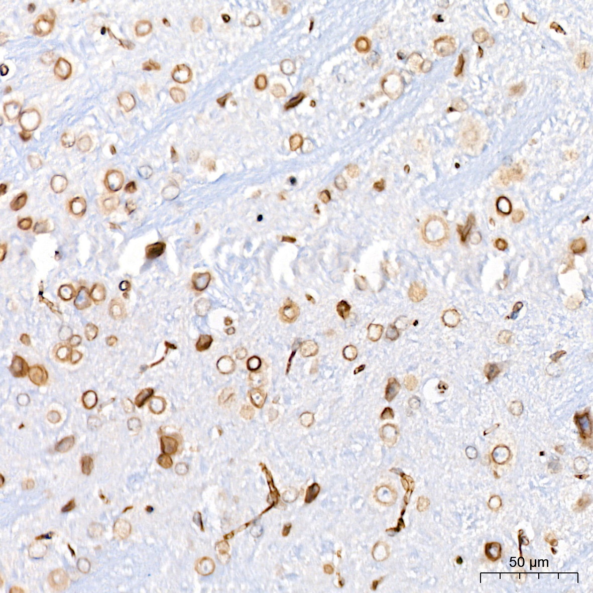
Immunohistochemistry analysis of paraffin-embedded Mouse brain tissue using SUN2 Rabbit mAb (CAB19782) at a dilution of 1:200 (40x lens). High pressure antigen retrieval performed with 0.01M Citrate buffer (pH 6.0) prior to IHC staining.


![Anti-SUN2 [R04-4M5] Monoclonal Antibody (AGMB00126)](https://cdn11.bigcommerce.com/s-h68l9z2lnx/images/stencil/590x590/products/271415/695030/anti-sun2-r04-4m5-monoclonal-antibody-agmb00126__78627.1774514359.jpg?c=2 "Anti-SUN2 [R04-4M5] Monoclonal Antibody (AGMB00126)")


