The ULK3 Monoclonal Antibody (CAB5959) is a high-quality antibody developed for reliable detection and analysis of target proteins. This antibody, generated in rabbits, exhibits high reactivity with human samples and has been validated for use in Western blot applications. It effectively binds to ULK3 protein, enabling precise detection and analysis in various cell types.ULK3 is a crucial player in the autophagy process, which is responsible for degrading and recycling cellular components to maintain cell health and functionality. Dysregulation of autophagy has been linked to various diseases, including cancer, neurodegenerative disorders, and metabolic conditions.
This antibody is validated for use in WB, IF/ICC, ELISA applications and has demonstrated reactivity against Human, Mouse samples.
Product Name:
ULK3 Monoclonal Antibody
SKU:
CAB5959
Size:
20μL, 100μL
Reactivity:
Human, Mouse
Clone Number:
ARC2118
Conjugate:
Unconjugated
Immunogen:
Synthetic peptide. This information is considered to be commercially sensitive.
Recommended starting concentration is 1 μg/mL. Please optimize the concentration based on your specific assay requirements.
Synonyms:
ULK3
Positive Sample:
293T
Cellular Localization:
Autophagosome, Ciliary Tip, Cytoplasm.
Calculated MW:
53kDa
Observed MW:
50kDa
Enables protein serine/threonine kinase activity. Involved in several processes, including fibroblast activation; protein autophosphorylation; and regulation of smoothened signaling pathway. Located in cytoplasm.
Purification Method
Affinity purification
Gene ID
25989
Buffer Information
Store at -20℃. Avoid freeze / thaw cycles. Buffer: PBS containing 50% glycerol and 0.05% BSA, preserved with proclin300 or sodium azide, pH 7.3.
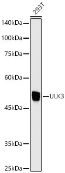
Western blot analysis of lysates from 293T cells using ULK3 Rabbit mAb (CAB5959) at 1:1000 dilution. Secondary antibody: HRP-conjugated Goat anti-Rabbit IgG (H+L) (CABS014) at 1:10000 dilution. Lysates/proteins: 25μg per lane. Blocking buffer: 3% nonfat dry milk in TBST. Detection: ECL Enhanced Kit (AbGn00021). Exposure time: 10s.
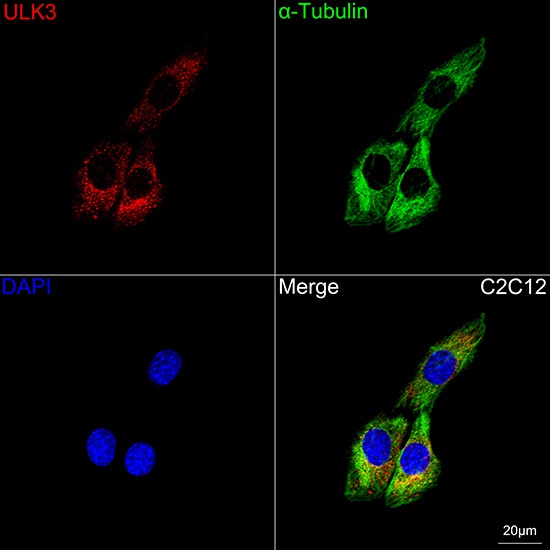
Confocal imaging of C2C12 cells using ULK3 Rabbit mAb (CAB5959, dilution 1:100) followed by a further incubation with Cy3 Goat Anti-Rabbit IgG (H+L) (CABS007, dilution 1:500) (Red). The cells were counterstained with α-Tubulin Mouse mAb (AC012, dilution 1:400) followed by incubation with ABflo® 488-conjugated Goat Anti-Mouse IgG (H+L) Ab (CABS076, dilution 1:500) (Green). DAPI was used for nuclear staining (Blue). Objective: 100x.