The AP1B1 Antibody (CAB16304) is a high-quality antibody developed for reliable detection and analysis of target proteins. This antibody, produced in rabbits, exhibits high reactivity with human samples and has been validated for use in Western blotting applications. By binding specifically to the AP1B1 protein, this antibody enables precise detection and analysis in a variety of cell types, making it an essential reagent for investigations in cell biology and molecular research.AP1B1 is a crucial component of the adaptor protein complex 1 (AP-1), which plays a critical role in the sorting of proteins to their proper destinations within the cell.
This antibody is validated for use in WB, ELISA applications and has demonstrated reactivity against Human, Mouse, Rat samples.
Product Name:
AP1B1 Antibody
SKU:
CAB16304
Size:
20μL, 100μL
Reactivity:
Human, Mouse, Rat
Conjugate:
Unconjugated
Immunogen:
Synthetic peptide. This information is considered to be commercially sensitive.
Recommended starting concentration is 1 μg/mL. Please optimize the concentration based on your specific assay requirements.
Synonyms:
ADTB1, BAM22, KIDAR, AP105A, CLAPB2, AP1B1
Positive Sample:
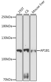
293T, C6, Mouse liver
Cellular Localization:
Cytosol, Early Endosome, Golgi Apparatus.
Calculated MW:
105kDa
Observed MW:
105kDa
Adaptor protein complex 1 is found at the cytoplasmic face of coated vesicles located at the Golgi complex, where it mediates both the recruitment of clathrin to the membrane and the recognition of sorting signals within the cytosolic tails of transmembrane receptors. This complex is a heterotetramer composed of two large, one medium, and one small adaptin subunit. The protein encoded by this gene serves as one of the large subunits of this complex and is a member of the adaptin protein family. This gene is a candidate meningioma gene. Alternative splicing results in multiple transcript variants.
Purification Method
Affinity purification
Gene ID
162
RRID
AB_2768382
Buffer Information
Store at -20℃. Avoid freeze / thaw cycles. Buffer: PBS with 0.01% thimerosal,50% glycerol,pH7.3.
Western blot analysis of various lysates using AP1B1 Rabbit pAb (CAB16304) at 1:1000 dilution. Secondary antibody: HRP-conjugated Goat anti-Rabbit IgG (H+L) (CABS014) at 1:10000 dilution. Lysates/proteins: 25μg per lane. Blocking buffer: 3% nonfat dry milk in TBST. Detection: ECL Basic Kit (AbGn00020). Exposure time: 5min.