The APOBEC3G Antibody (CAB1459) is a high-quality antibody developed for reliable detection and analysis of target proteins. This antibody, produced in rabbits, is highly specific to human APOBEC3G and has been validated for use in Western blotting applications. By binding to the APOBEC3G protein, this antibody allows for accurate detection and analysis in various cell types, making it ideal for studies in virology, immunology, and cancer research.
This antibody is validated for use in WB, IHC-P, ELISA applications and has demonstrated reactivity against Human, Mouse, Rat samples.
Product Name:
APOBEC3G Antibody
SKU:
CAB1459
Size:
20μL, 100μL
Reactivity:
Human, Mouse, Rat
Conjugate:
Unconjugated
Immunogen:
Recombinant protein (or fragment).This information is considered to be commercially sensitive.
This gene is a member of the cytidine deaminase gene family. It is one of seven related genes or pseudogenes found in a cluster, thought to result from gene duplication, on chromosome 22. Members of the cluster encode proteins that are structurally and functionally related to the C to U RNA-editing cytidine deaminase APOBEC1. The protein encoded by this gene catalyzes site-specific deamination of both RNA and single-stranded DNA. The encoded protein has been found to be a specific inhibitor of human immunodeficiency virus-1 (HIV-1) infectivity.
Purification Method
Affinity purification
Gene ID
60489
RRID
AB_2761464
Buffer Information
Store at -20℃. Avoid freeze / thaw cycles. Buffer: PBS with 0.09% Sodium azide,50% glycerol,pH7.3.
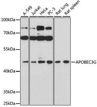
Western blot analysis of various lysates using APOBEC3G Rabbit pAb (CAB1459) at 1:1000 dilution. Secondary antibody: HRP-conjugated Goat anti-Rabbit IgG (H+L) (CABS014) at 1:10000 dilution. Lysates/proteins: 25μg per lane. Blocking buffer: 3% nonfat dry milk in TBST. Detection: ECL Basic Kit (AbGn00020). Exposure time: 3min.
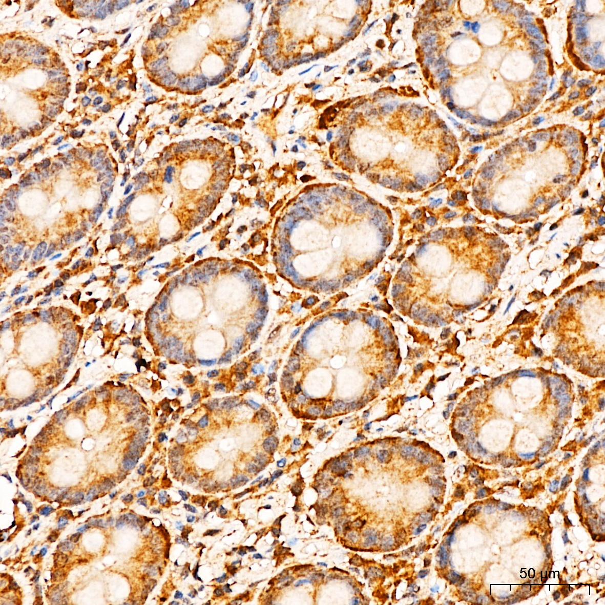
Immunohistochemistry analysis of paraffin-embedded Human colon tissue using APOBEC3G Rabbit pAb (CAB1459) at a dilution of 1:300 (40x lens). High pressure antigen retrieval performed with 0.01M Citrate buffer (pH 6.0) prior to IHC staining.
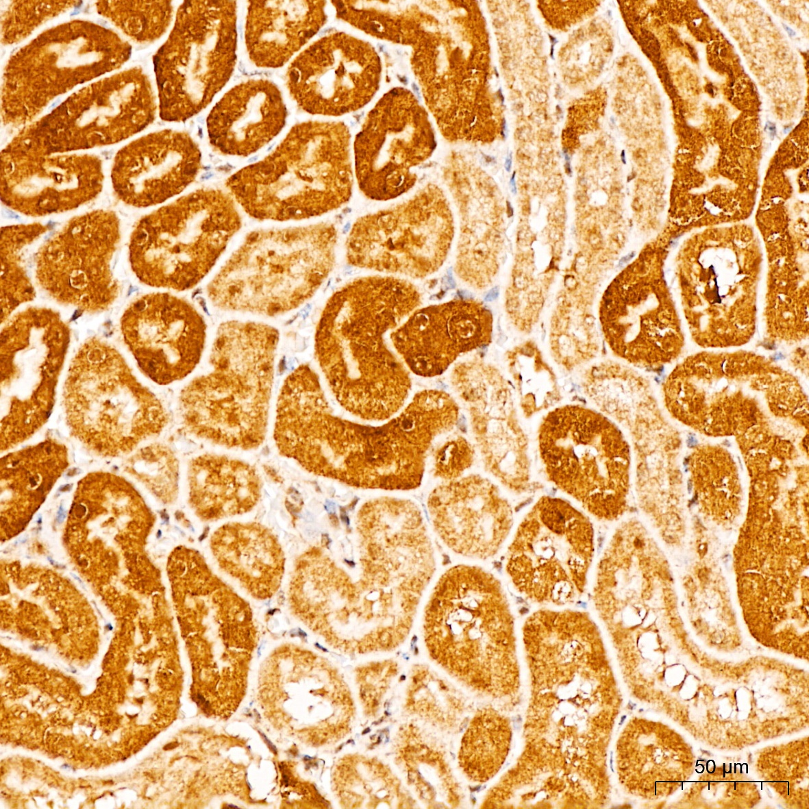
Immunohistochemistry analysis of paraffin-embedded Mouse kidney tissue using APOBEC3G Rabbit pAb (CAB1459) at a dilution of 1:300 (40x lens). High pressure antigen retrieval performed with 0.01M Citrate buffer (pH 6.0) prior to IHC staining.