The ASAH1 Antibody (CAB13948) is a high-quality antibody developed for reliable detection and analysis of target proteins. This antibody, produced in rabbits, has high specificity for human samples and has been validated for use in Western blot applications. By binding to the ASAH1 protein, researchers can accurately detect and analyze the expression of ASAH1 in a variety of cell types, making it ideal for studies in lipid metabolism, signal transduction, and cancer research.
This antibody is validated for use in WB, IF/ICC, ELISA applications and has demonstrated reactivity against Human, Mouse, Rat samples.
Product Name:
ASAH1 Antibody
SKU:
CAB13948
Size:
20μL, 100μL
Reactivity:
Human, Mouse, Rat
Conjugate:
Unconjugated
Immunogen:
Recombinant protein (or fragment).This information is considered to be commercially sensitive.
Recommended starting concentration is 1 μg/mL. Please optimize the concentration based on your specific assay requirements.
Synonyms:
AC, PHP, ASAH, PHP32, ACDase, SMAPME, ASAH1
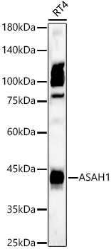
Positive Sample:
RT4
Cellular Localization:
Lysosome.
Calculated MW:
45kDa
Observed MW:
40kDa
This gene encodes a member of the acid ceramidase family of proteins. Alternative splicing results in multiple transcript variants, at least one of which encodes a preproprotein that is proteolytically processed. Processing of this preproprotein generates alpha and beta subunits that heterodimerize to form the mature lysosomal enzyme, which catalyzes the degradation of ceramide into sphingosine and free fatty acid. This enzyme is overexpressed in multiple human cancers and may play a role in cancer progression. Mutations in this gene are associated with the lysosomal storage disorder, Farber lipogranulomatosis, and a neuromuscular disorder, spinal muscular atrophy with progressive myoclonic epilepsy.
Purification Method
Affinity purification
Gene ID
427
RRID
AB_2760801
Buffer Information
Store at -20℃. Avoid freeze / thaw cycles. Buffer: PBS containing 50% glycerol, preserved with proclin300 or sodium azide, pH 7.3.
Western blot analysis of lysates from RT4 cells using ASAH1 Rabbit pAb (CAB13948) at 1:700 dilution. Secondary antibody: HRP-conjugated Goat anti-Rabbit IgG (H+L) (CABS014) at 1:10000 dilution. Lysates/proteins: 25 μg per lane. Blocking buffer: 3% nonfat dry milk in TBST. Detection: ECL Basic Kit (AbGn00020). Exposure time: 60s.
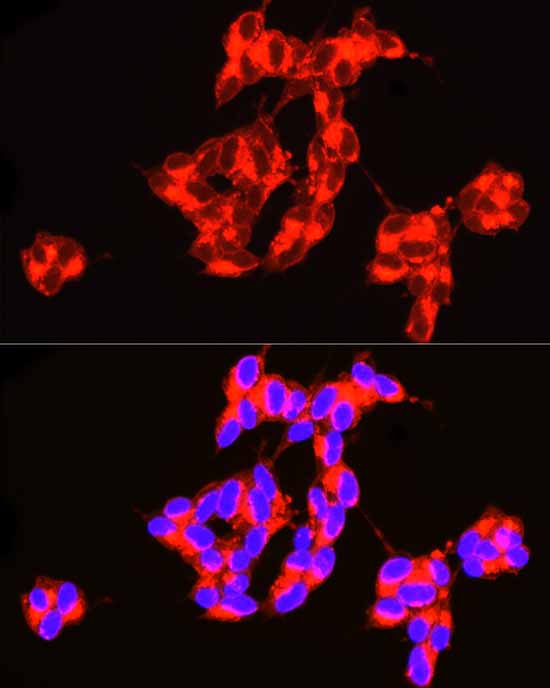
Immunofluorescence analysis of SH-SY5Y cells using ASAH1 Rabbit pAb (CAB13948) at dilution of 1:100 (40x lens). Secondary antibody: Cy3-conjugated Goat anti-Rabbit IgG (H+L) (CABS007) at 1:500 dilution. Blue: DAPI for nuclear staining.