The ATG7 Monoclonal Antibody (CAB19604) is a high-quality antibody developed for reliable detection and analysis of target proteins. This antibody, developed using rabbit monoclonal technology, exhibits high specificity and sensitivity towards ATG7 protein in human samples.ATG7 is a key component of the autophagy pathway, responsible for the conjugation of ATG8/LC3 to phosphatidylethanolamine and promoting the formation of autophagosomes. Dysregulation of autophagy has been linked to various diseases, including cancer, neurodegenerative disorders, and metabolic conditions, making ATG7 an important target for therapeutic interventions.
This antibody is validated for use in WB, IHC-P, IP, ELISA applications and has demonstrated reactivity against Human, Mouse, Rat samples.
Product Name:
ATG7 Monoclonal Antibody
SKU:
CAB19604
Size:
20μL, 100μL
Reactivity:
Human, Mouse, Rat
Clone Number:
ARC0083
Conjugate:
Unconjugated
Immunogen:
Synthetic peptide. This information is considered to be commercially sensitive.
0.5μg-4μg antibody for 200μg-400μg extracts of whole cells
ELISA
Recommended starting concentration is 1 μg/mL. Please optimize the concentration based on your specific assay requirements.
Synonyms:
GSA7, [KD Validated] ATG7
Positive Sample:
HeLa, THP-1, 293T, Mouse brain, Mouse lung, Mouse spleen, Rat testis, HeLa
Cellular Localization:
Cytoplasm, Preautophagosomal Structure.
Calculated MW:
78kDa
Observed MW:
78kDa
This gene encodes an E1-like activating enzyme that is essential for autophagy and cytoplasmic to vacuole transport. The encoded protein is also thought to modulate p53-dependent cell cycle pathways during prolonged metabolic stress. It has been associated with multiple functions, including axon membrane trafficking, axonal homeostasis, mitophagy, adipose differentiation, and hematopoietic stem cell maintenance. Alternative splicing results in multiple transcript variants.
Purification Method
Affinity purification
Gene ID
10533
RRID
AB_2862693
Buffer Information
Store at -20℃. Avoid freeze / thaw cycles. Buffer: PBS containing 50% glycerol and 0.05% BSA, preserved with proclin300 or sodium azide, pH 7.3.
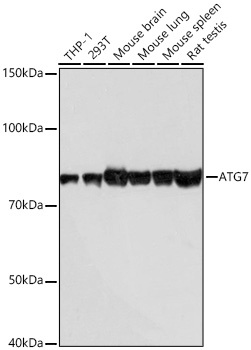
Western blot analysis of various lysates using [KD Validated] ATG7 Rabbit mAb (CAB19604) at 1:1000 dilution incubated overnight at 4℃. Secondary antibody: HRP-conjugated Goat anti-Rabbit IgG (H+L) (CABS014) at 1:10000 dilution. Lysates/proteins: 25μg per lane. Blocking buffer: 3% nonfat dry milk in TBST. Detection: ECL Basic Kit (AbGn00020). Exposure time: 10s.
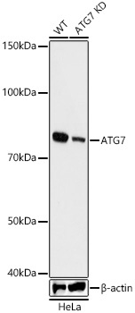
Western blot analysis of lysates from wild type(WT) and ATG7 knockdown (KD) HeLa cells using [KD Validated] ATG7 Rabbit mAb (CAB19604) at 1:1000 dilution incubated overnight at 4℃. Secondary antibody: HRP-conjugated Goat anti-Rabbit IgG (H+L) (CABS014) at 1:10000 dilution. Lysates/proteins: 25μg per lane. Blocking buffer: 3% nonfat dry milk in TBST. Detection: ECL Basic Kit (AbGn00020). Exposure time: 60s.
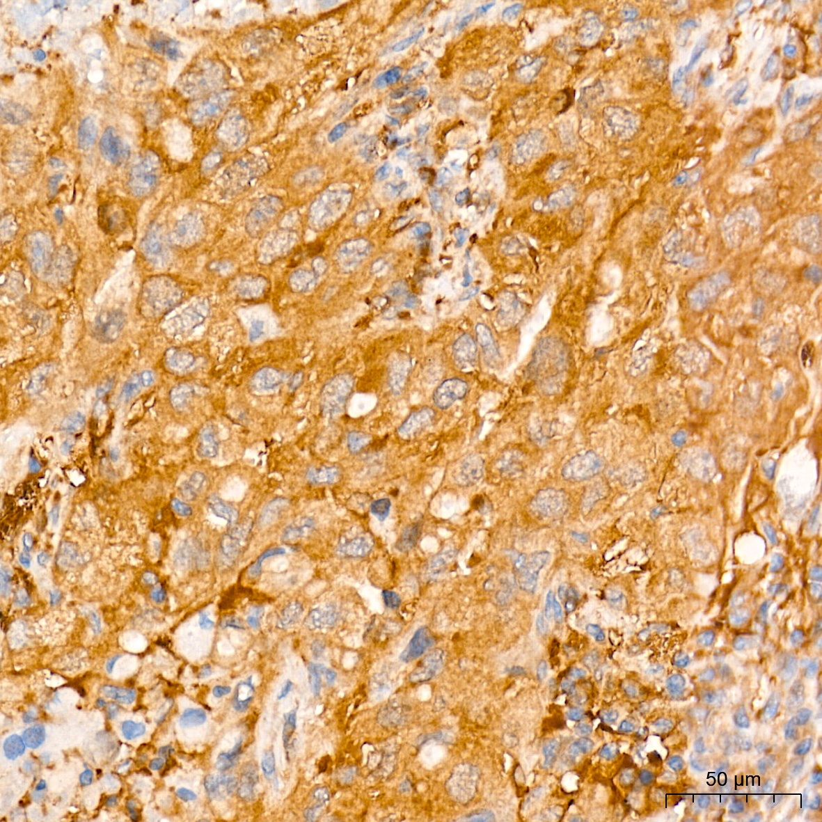
Immunohistochemistry analysis of paraffin-embedded Human lung cancer tissue using [KD Validated] ATG7 Rabbit mAb (CAB19604) at a dilution of 1:500 (40x lens). High pressure antigen retrieval performed with 0.01M Tris-EDTA Buffer (pH 9.0) prior to IHC staining.
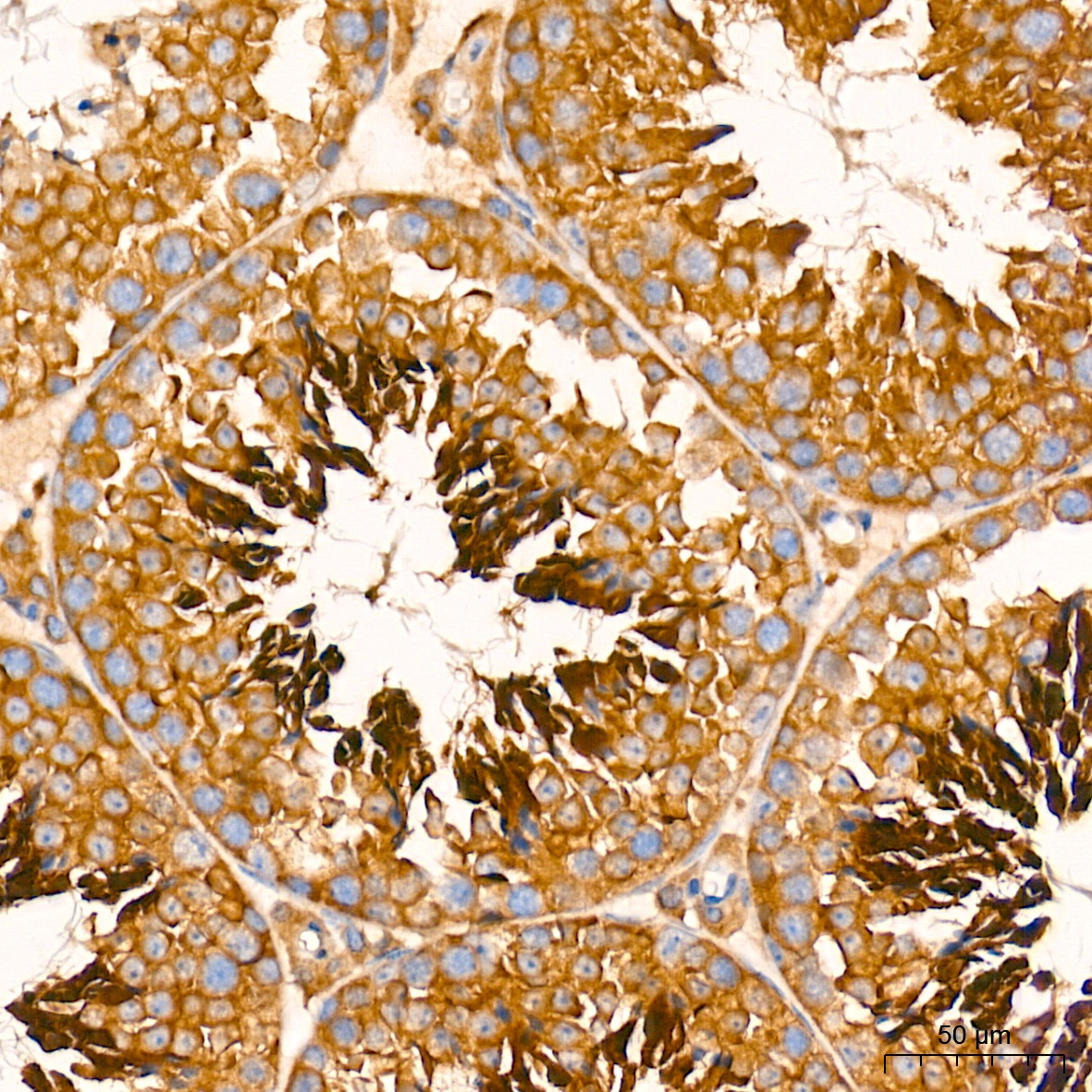
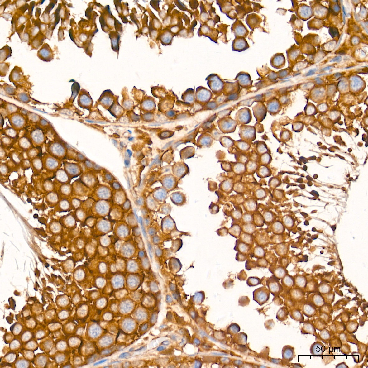
Immunohistochemistry analysis of paraffin-embedded Mouse testis tissue using [KD Validated] ATG7 Rabbit mAb (CAB19604) at a dilution of 1:500 (40x lens). High pressure antigen retrieval performed with 0.01M Tris-EDTA Buffer (pH 9.0) prior to IHC staining.
Immunohistochemistry analysis of paraffin-embedded Rat testis tissue using [KD Validated] ATG7 Rabbit mAb (CAB19604) at a dilution of 1:500 (40x lens). High pressure antigen retrieval performed with 0.01M Tris-EDTA Buffer (pH 9.0) prior to IHC staining.
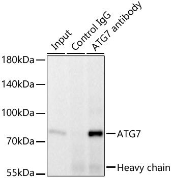
Immunoprecipitation of ATG7 from 300 µg extracts of HeLa cells was performed using 2 µg of [KD Validated] ATG7 Rabbit mAb (CAB19604). Rabbit IgG isotype control (AC005) was used to precipitate the Control IgG sample. IP samples were eluted with 1X reducing Laemmli Buffer. The Input lane represents 10% of the total input. Western blot analysis of immunoprecipitates was conducted using [KD Validated] ATG7 Rabbit mAb (CAB19604) at a dilution of 1:1000.