The ATPIF1 Antibody (CAB5099) is a high-quality antibody developed for reliable detection and analysis of target proteins. The antibody, raised in rabbits, is highly reactive with human samples and is validated for use in Western blot applications. It specifically binds to the ATP5F1 protein, allowing for precise detection and analysis in various cell types, making it an essential tool for studies in biochemistry and cell biology.ATP5F1, also known as ATP synthase F1 subunit alpha, plays a crucial role in the generation of ATP, the energy currency of the cell. Its involvement in ATP synthesis makes it a vital component of cellular metabolism and energy production processes.
This antibody is validated for use in WB, IF/ICC, IP, ELISA applications and has demonstrated reactivity against Human, Mouse samples.
Product Name:
ATPIF1 Antibody
SKU:
CAB5099
Size:
20μL, 100μL
Reactivity:
Human, Mouse
Conjugate:
Unconjugated
Immunogen:
Recombinant protein (or fragment).This information is considered to be commercially sensitive.
0.5μg-4μg antibody for 200μg-400μg extracts of whole cells
ELISA
Recommended starting concentration is 1 μg/mL. Please optimize the concentration based on your specific assay requirements.
Synonyms:
IP, ATPI, ATPIP, ATPIF1, F1
Positive Sample:
293T
Cellular Localization:
Mitochondrion.
Calculated MW:
12kDa
Observed MW:
12kDa
Enables several functions, including ATPase binding activity; angiostatin binding activity; and mitochondrial proton-transporting ATP synthase complex binding activity. Involved in several processes, including mitochondrial depolarization; negative regulation of ATPase activity; and regulation of protein targeting to mitochondrion. Located in cell surface and mitochondrion.
Purification Method
Affinity purification
Gene ID
93974
RRID
AB_2766016
Buffer Information
Store at -20℃. Avoid freeze / thaw cycles. Buffer: PBS containing 50% glycerol, preserved with proclin300 or sodium azide, pH 7.3.
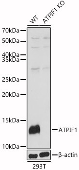
Western blot analysis of lysates from wild type(WT) and ATPIF1 knockout (KO) 293T(KO) cells, using [KO Validated] ATPIF1 Rabbit pAb (CAB5099) at 1:1000 dilution. Secondary antibody: HRP-conjugated Goat anti-Rabbit IgG (H+L) (CABS014) at 1:10000 dilution. Lysates/proteins: 25μg per lane. Blocking buffer: 3% nonfat dry milk in TBST. Detection: ECL Basic Kit (AbGn00020). Exposure time: 1s.
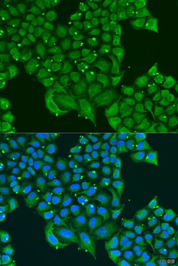
Immunofluorescence analysis of U2OS cells using [KO Validated] ATPIF1 Rabbit pAb (CAB5099) at dilution of 1:100. Secondary antibody: Cy3-conjugated Goat anti-Rabbit IgG (H+L) (CABS007) at 1:500 dilution. Blue: DAPI for nuclear staining.