


Description
[KO Validated] Bax Antibody (CAB0207)
The [KO Validated] Bax Antibody (CAB0207) is a high-quality antibody developed for reliable detection and analysis of target proteins. This antibody, produced in rabbits, exhibits high reactivity with human samples and has been validated for use in Western blot applications.Bax is a pro-apoptotic protein that plays a critical role in the intrinsic pathway of apoptosis, promoting cell death under various conditions such as DNA damage or cellular stress. Dysregulation of Bax can have profound effects on cell survival and contribute to the development of diseases such as cancer.With the ability to specifically bind to Bax, this antibody enables researchers to detect and analyze the expression and localization of the protein in different cell types.
This antibody is validated for use in WB, IHC-P, IF/ICC, IP, ELISA applications and has demonstrated reactivity against Human, Mouse, Rat samples.
| Product Name: | [KO Validated] Bax Antibody |
| SKU: | CAB0207 |
| Size: | 20μL, 100μL |
| Reactivity: | Human, Mouse, Rat |
| Conjugate: | Unconjugated |
| Immunogen: | Synthetic peptide. This information is considered to be commercially sensitive. | ||||||||||
| Sequence: | MDGS GEQP RGGG PTSS EQIM KTGA LLLQ GFIQ DRAG RMGG EAPE LALD PVPQ DAST KKLS ECLK RIGD ELDS NMEL QRMI AAVD TDSP REVF FRVA ADMF | ||||||||||
| Tested Applications: | WB IHC-P IF/ICC IP ELISA | ||||||||||
| Recommended Dilution: |
| ||||||||||
| Synonyms: | BCL2 Associated X, Bcl-2-Like Protein 4, Bcl2-L-4, BCL2L4, BAX |
| Positive Sample: | MCF7, NIH/3T3, Mouse lung, PC-12, 293T |
| Cellular Localization: | Cytoplasm, Cytoplasm, Mitochondrion Membrane, Single-Pass Membrane Protein. |
| Calculated MW: | 21kDa |
| Observed MW: | 21kDa |
BAX (also known as BCL2 Associated X, Bcl-2-Like Protein 4, Bcl2-L-4, BCL2L4) is a member of the BCL2 family of proteins that play a key role in the regulation of apoptosis in higher eukaryotes (https://www.uniprot.org/uniprot/Q07812). BAX comprises 4 Bcl-2 homology domains (BH1-BH4) and a C-terminal transmembrane domain. In healthy mammalian cells, BAX is localized to the cytoplasm through its interaction with the anti-apoptotic BL-2 family members BCL2L1/Bcl-xL . In response to apoptotic stimuli, however, BAX undergoes a conformational change that causes it to translocate to the outer mitochondrial membrane where it initiates the mitochondrial pathway of apoptosis via two potential mechanisms. Firstly, upon translocation to the outer mitochondrial membrane, BAX interacts with the mitochondrial voltage-dependent anion channel (VDAC) leading to the opening of the channel, loss of membrane potential, and the release of cytochrome c from the mitochondrion . The release of cytochrome C into the cytoplasm leads to the activation of Caspase3, initiating apoptosis. Secondly, activated BAX forms homodimers, which then assemble into oligomers on the mitochondrial outer membrane to create pores that permeabilize the mitochondrion leading to the release of cytochrome C.BAX has been shown to be involved in p53-mediated apoptosis. Expression of the human bax gene has been shown to be directly regulated by p53, and the bax promoter contains four motifs with homology to consensus p53-binding sites. Furthermore, p53 directly interacts with BAX to promote its activation.
| Purification Method | Affinity purification |
| Gene ID | 581 |
| RRID | AB_2757021 |
| Buffer Information | Store at -20℃. Avoid freeze / thaw cycles. Buffer: PBS with 0.09% Sodium azide,50% glycerol,pH7.3. |
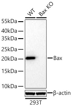 | Western blot analysis of lysates from wild type (WT) and Bax knockout (KO) 293T cells using [KO Validated] Bax Rabbit pAb (CAB0207) at 1:3000 dilution incubated at room temperature for 1.5 hours. Secondary antibody: HRP-conjugated Goat anti-Rabbit IgG (H+L) (CABS014) at 1:10000 dilution. Lysates/proteins: 25 μg per lane. Blocking buffer: 3% nonfat dry milk in TBST. Detection: ECL Basic Kit (AbGn00020). Exposure time: 5 s. |
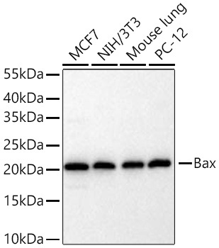 | Western blot analysis of various lysates using [KO Validated] Bax Rabbit pAb (CAB0207) at 1:3000 dilution incubated at room temperature for 1.5 hours. Secondary antibody: HRP-conjugated Goat anti-Rabbit IgG (H+L) (CABS014) at 1:10000 dilution. Lysates/proteins: 25 μg per lane. Blocking buffer: 3% nonfat dry milk in TBST. Detection: ECL Basic Kit (AbGn00020). Exposure time: 5 s. |
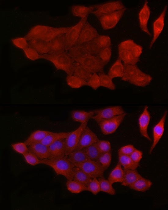 | Immunofluorescence analysis of HeLa cells using [KO Validated] Bax Rabbit pAb (CAB0207) at dilution of 1:50 (40x lens). Secondary antibody: Cy3-conjugated Goat anti-Rabbit IgG (H+L) (CABS007) at 1:500 dilution. Blue: DAPI for nuclear staining. |
 | Immunofluorescence analysis of HepG2 cells using [KO Validated] Bax Rabbit pAb (CAB0207) at dilution of 1:50 (40x lens). Secondary antibody: Cy3-conjugated Goat anti-Rabbit IgG (H+L) (CABS007) at 1:500 dilution. Blue: DAPI for nuclear staining. |
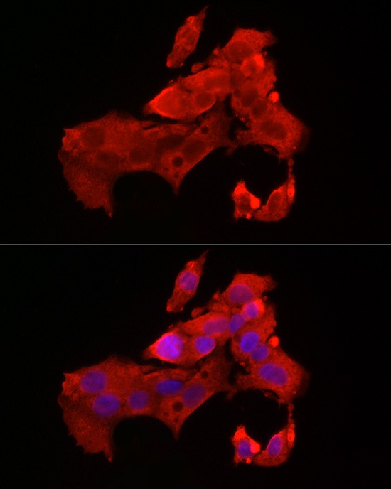
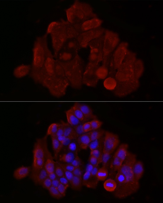 | Immunofluorescence analysis of MCF7 cells using [KO Validated] Bax Rabbit pAb (CAB0207) at dilution of 1:50 (40x lens). Secondary antibody: Cy3-conjugated Goat anti-Rabbit IgG (H+L) (CABS007) at 1:500 dilution. Blue: DAPI for nuclear staining. |
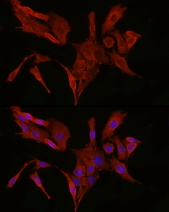 | Immunofluorescence analysis of PC-12 cells using [KO Validated] Bax Rabbit pAb (CAB0207) at dilution of 1:50 (40x lens). Secondary antibody: Cy3-conjugated Goat anti-Rabbit IgG (H+L) (CABS007) at 1:500 dilution. Blue: DAPI for nuclear staining. |


