The BCKDK Antibody (CAB8184) is a high-quality antibody developed for reliable detection and analysis of target proteins. This antibody, generated in rabbits, is highly specific to human samples and is validated for use in Western blot applications. By binding to the BCKDK protein, this antibody allows for the detection and analysis of BCKDK in a variety of cell types, making it an essential component of studies in metabolism and metabolic disorders.BCKDK, also known as branched-chain ketoacid dehydrogenase kinase, is involved in regulating the activity of the branched-chain ketoacid dehydrogenase complex, which is responsible for breaking down branched-chain amino acids.
This antibody is validated for use in WB, IF/ICC, ELISA applications and has demonstrated reactivity against Human, Rat samples.
Product Name:
BCKDK Antibody
SKU:
CAB8184
Size:
20μL, 100μL
Reactivity:
Human, Rat
Conjugate:
Unconjugated
Immunogen:
Recombinant protein (or fragment).This information is considered to be commercially sensitive.
Recommended starting concentration is 1 μg/mL. Please optimize the concentration based on your specific assay requirements.
Synonyms:
BDK, BCKDKD, BCKDK
Positive Sample:
22Rv1
Cellular Localization:
Mitochondrion, Mitochondrion Matrix.
Calculated MW:
46kDa
Observed MW:
46kDa
The branched-chain alpha-ketoacid dehydrogenase complex (BCKD) is an important regulator of the valine, leucine, and isoleucine catabolic pathways. The protein encoded by this gene is found in the mitochondrion, where it phosphorylates and inactivates BCKD. Several transcript variants encoding different isoforms have been found for this gene.
Purification Method
Affinity purification
Gene ID
10295
RRID
AB_2768578
Buffer Information
Store at -20℃. Avoid freeze / thaw cycles. Buffer: PBS containing 50% glycerol, preserved with proclin300 or sodium azide, pH 7.3.
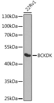
Western blot analysis of lysates from 22Rv1 cells, using BCKDK Rabbit pAb (CAB8184) at 1:1000 dilution. Secondary antibody: HRP-conjugated Goat anti-Rabbit IgG (H+L) (CABS014) at 1:10000 dilution. Lysates/proteins: 25μg per lane. Blocking buffer: 3% nonfat dry milk in TBST. Detection: ECL Basic Kit (AbGn00020). Exposure time: 90s.
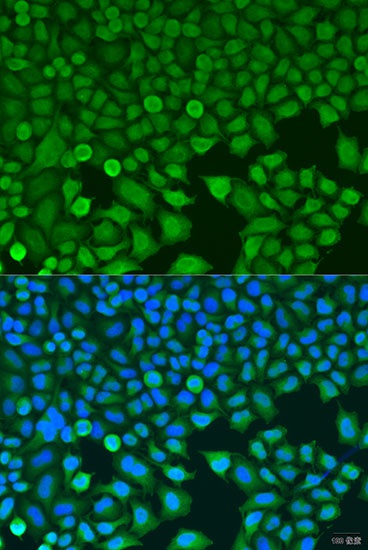
Immunofluorescence analysis of U2OS cells using BCKDK Rabbit pAb (CAB8184) at dilution of 1:100. Secondary antibody: Cy3-conjugated Goat anti-Rabbit IgG (H+L) (CABS007) at 1:500 dilution. Blue: DAPI for nuclear staining.


 at 1:10000 dilution. Lysates/proteins: 25ug per lane. Blocking buffer: 3% nonfat dry milk in TBST. Detection: ECL Basic Kit. Exposure time: 30s.")


