
Cleaved Caspase-3 and Apoptosis
Cleaved Caspase-3: Activation, Function and Detection in Apoptosis
Caspase-3 sits at the point where apoptotic signalling becomes irreversible. It is made as an inactive proenzyme and only becomes competent once cleaved by an initiator caspase — which is why the cleaved form, not total caspase-3, is the marker of apoptosis. This guide covers its structure and activation, the DEVD specificity that underpins every caspase-3 assay, how it is regulated, and how to detect it without over-reading the result.
Browse caspase reagents →Key takeaways
- Caspase-3 is synthesised as an inactive 32 kDa proenzyme and becomes active only after cleavage into large and small subunits, which assemble into a heterotetramer.
- Because activation is proteolytic rather than transcriptional, total caspase-3 barely changes during apoptosis — only the cleaved form reports activity.
- Caspase-3 is cleaved by initiator caspase-8 (extrinsic pathway) or caspase-9 (intrinsic pathway), which is why measuring the initiators tells you which route was taken.
- It cleaves substrates after aspartate in a DEVD motif, and that specificity is the basis of every fluorogenic caspase-3/7 activity assay.
- Cleaved PARP is the classic downstream confirmation that active caspase-3 was doing something, not merely present.
- Regulation runs through IAPs such as XIAP and survivin, opposed by Smac/DIABLO, and upstream through the Bcl-2 family balance controlling cytochrome c release.
- Caspase-3 also has non-apoptotic roles — in differentiation, neuronal pruning and tumour repopulation — so its presence does not always mean a cell is dying.
Reagents for caspase-3 and apoptosis
The panel below follows the cascade — both initiator caspases, the executioner in active and total form, a functional activity assay, and the substrate that confirms cleavage occurred.
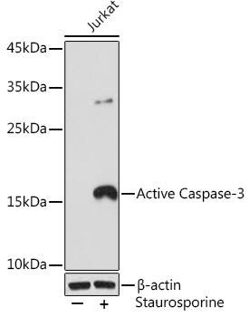
Active Caspase-3 Rabbit Monoclonal Antibody
Recognises the neo-epitope exposed only after cleavage — detects active caspase-3 without cross-reacting with the proenzyme.
View product →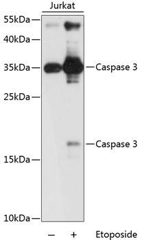
Caspase-3 Rabbit Polyclonal Antibody (KO validated)
Total caspase-3 including the proenzyme — the control that shows a rise in cleaved form is not a rise in expression.
View product →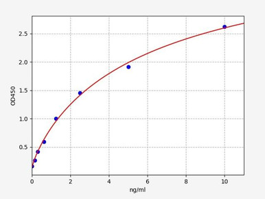
Human Caspase-8 ELISA Kit
The extrinsic-pathway initiator; measuring it indicates death-receptor signalling upstream.
View product →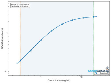
Human Caspase-9 ELISA Kit
The intrinsic-pathway initiator, activated in the apoptosome after cytochrome c release.
View product →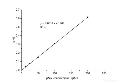
Caspase-3/7 Activity Assay Kit
Uses a DEVD-based substrate to measure enzymatic activity rather than protein presence.
View product →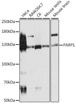
PARP1 Rabbit Polyclonal Antibody (KO validated)
PARP1 is the canonical caspase-3 substrate — its cleavage confirms the enzyme was functionally active.
View product →What are caspases?
Caspases are cysteine-dependent aspartate-specific proteases — the name compresses both properties. They use a catalytic cysteine to cleave their substrates, and they cut specifically after an aspartate residue, which is unusual among proteases and gives the family its precision.
They divide by function. Initiator caspases — caspase-8, -9 and -10 — sit at the top of the cascade, are activated by recruitment into signalling complexes, and their job is to cleave other caspases. Executioner caspases — caspase-3, -6 and -7 — are activated by the initiators and cleave the several hundred cellular substrates whose destruction constitutes apoptosis. A separate group, the inflammatory caspases including caspase-1, -4 and -5, serve pyroptosis and cytokine maturation rather than apoptosis.
Caspase-3 is the principal executioner. Caspase-7 overlaps with it substantially and shares the same substrate preference, so the two are usually measured together — which is why activity assays are labelled caspase-3/7 rather than caspase-3 alone.
Caspase-3 structure
Caspase-3 is a 277-amino-acid protein, produced as an inactive proenzyme of roughly 32 kDa called procaspase-3. The proenzyme comprises an N-terminal prodomain followed by the sequences that will become the large and small subunits, separated by cleavable linkers.
Activation cleaves the proenzyme into a large subunit of about 17 kDa and a small subunit of about 12 kDa, with the short prodomain removed. Two large and two small subunits then assemble into a heterotetramer — the active enzyme is not a simple pair of subunits but a dimer of heterodimers, with two independent active sites.
Each active site sits at the interface between a large and a small subunit and contains the catalytic cysteine, together with a pocket that reads the four residues preceding the cleavage point. In the proenzyme that pocket is misaligned, which is why procaspase-3 has essentially no catalytic activity — activation is a structural rearrangement, not the removal of a plug.
Activation and the cleaved form
Procaspase-3 is cleaved by an initiator caspase, and which initiator does it identifies the upstream pathway.
The extrinsic pathway
Death ligands such as TNF-alpha, FasL and TRAIL bind their receptors, which recruit adaptor proteins and procaspase-8 into a death-inducing signalling complex. Clustering activates caspase-8, which then cleaves procaspase-3 directly. In some cell types caspase-8 also cleaves Bid, feeding into the mitochondrial pathway and amplifying the signal.
The intrinsic pathway
Cellular stress — DNA damage, growth factor withdrawal, hypoxia — shifts the Bcl-2 family balance so that Bax and Bak permeabilise the outer mitochondrial membrane. Cytochrome c escapes into the cytosol and binds Apaf-1, which oligomerises with procaspase-9 into the apoptosome, a wheel-shaped complex that activates caspase-9. Caspase-9 then cleaves procaspase-3.
Both routes converge on the same step, which is why caspase-3 is described as the point of convergence and, once cleaved, the point of no return. The cell can still be rescued upstream of caspase-3 activation; after it, dismantling proceeds.
Substrate specificity and DEVD
Caspase-3 recognises a four-residue motif immediately N-terminal to the cleavage site and cuts after the aspartate. Its preferred motif is DEVD — Asp-Glu-Val-Asp — with cleavage occurring after the final aspartate.
This is not merely a structural footnote; it is the basis of the entire measurement toolkit. Fluorogenic and colorimetric caspase-3 assays use synthetic DEVD peptides coupled to a reporter, so that cleavage releases signal in proportion to enzymatic activity. It is also why those assays cannot distinguish caspase-3 from caspase-7, which shares the preference — hence the caspase-3/7 labelling.
Among several hundred known substrates, a few are worth naming because they explain the morphology of apoptosis. PARP1 is cleaved, disabling DNA repair. ICAD is cleaved, releasing the nuclease CAD to fragment DNA — the origin of the DNA laddering that TUNEL detects. Lamins are cleaved, causing nuclear collapse. ROCK1 is cleaved, driving the membrane blebbing characteristic of apoptotic cells. Each visible feature of apoptosis traces back to a specific cleavage event.
Role in the execution phase
Once active, cleaved caspase-3 dismantles the cell in a controlled sequence rather than destroying it indiscriminately.
- Structural breakdown. Cleavage of cytoskeletal and nuclear lamina proteins collapses cell and nuclear architecture, producing the shrinkage and nuclear condensation seen histologically.
- DNA fragmentation. By cleaving ICAD, caspase-3 releases CAD to cut chromatin between nucleosomes, generating the characteristic ladder of fragments.
- Disabling repair and replication. PARP1 cleavage removes DNA repair capacity, and cleavage of replication and transcription machinery halts the cell’s ongoing functions.
- Signalling for clearance. Caspase-3-dependent events lead to phosphatidylserine exposure on the outer membrane, the signal phagocytes recognise. This is why apoptosis is non-inflammatory: contents stay membrane-bound and are cleared before rupture, in contrast to necrosis where membrane failure releases them.
- Membrane blebbing. ROCK1 cleavage drives the formation of apoptotic bodies, packaging the remains for phagocytosis.
Regulation of activation
Because caspase-3 activation is effectively irreversible, it is restrained at several levels.
| Regulator | Effect | Mechanism |
|---|---|---|
| XIAP | Inhibits | Binds and directly blocks the active sites of caspase-3 and -7; the most potent endogenous caspase inhibitor |
| Survivin | Inhibits | IAP family member, frequently overexpressed in tumours; also functions in mitosis |
| Smac/DIABLO | Promotes | Released from mitochondria during apoptosis and sequesters IAPs, freeing caspase-3 |
| cIAP1 and cIAP2 | Modulate | Ubiquitin ligases that regulate upstream death-receptor signalling and caspase turnover |
| Bcl-2 and Bcl-xL | Inhibit | Prevent Bax and Bak pore formation, blocking cytochrome c release and so preventing caspase-9 activation |
| Bax, Bak, Bid, Bim | Promote | Permeabilise the outer mitochondrial membrane, initiating the intrinsic route |
The therapeutic relevance follows directly. Smac mimetics were developed to neutralise IAPs and lower the threshold for caspase activation, and BH3 mimetics such as venetoclax block Bcl-2 to release Bax and Bak. Both work by shifting these balances rather than by acting on caspase-3 itself, which has proved difficult to target directly.
Detecting cleaved caspase-3
This is where most interpretive errors arise, so it is worth being precise about what each method reports.
| Method | What it reports | Caveat |
|---|---|---|
| Cleaved-specific antibody | Presence of the active form, via a neo-epitope exposed only after cleavage | Detects protein, not activity — an inhibited enzyme still stains positive |
| Total caspase-3 antibody | Proenzyme plus cleaved form | Barely changes during apoptosis; useful only as a loading and expression control |
| DEVD activity assay | Enzymatic activity in the sample | Cannot distinguish caspase-3 from caspase-7 |
| Cleaved PARP antibody | That active caspase-3 cleaved a real substrate | Indirect — other proteases can contribute |
| TUNEL | DNA fragmentation, downstream of caspase-3 | Late-stage; also positive in some necrotic cells |
Why the antibody must be cleavage-specific
Cleavage exposes a new N-terminus on the large subunit that does not exist in the proenzyme. Antibodies raised against that junction bind only the processed form, which is what makes them informative — a total caspase-3 antibody stains both and will look unchanged whether or not apoptosis is occurring.
Practical guidance
- Run cleaved and total together. A rise in cleaved signal means little unless total is unchanged, exactly as with phospho-specific work.
- Pair protein detection with activity. Cleaved caspase-3 can be present yet held inactive by XIAP, so an antibody-positive sample is not proof of enzymatic activity. A DEVD assay closes that gap.
- Confirm with a substrate. Cleaved PARP demonstrates the enzyme acted, which is stronger evidence than the enzyme being present.
- Take a time course. Caspase-3 activation is transient — cells that have already fragmented and been cleared no longer stain, so a late single timepoint can substantially underestimate apoptosis.
- Include phosphatase and protease inhibitors, and keep lysates cold. Caspase activity in lysate can continue and generate cleavage artefacts.
Non-apoptotic roles
An important caveat for anyone using cleaved caspase-3 as an apoptosis marker: the enzyme has functions that do not kill the cell. Limited, localised caspase-3 activity contributes to skeletal muscle and bone differentiation, to neuronal pruning and synaptic remodelling during development, and to erythroid maturation.
Most consequentially for cancer research, caspase-3 activity in dying tumour cells can promote proliferation of surviving neighbours, through release of prostaglandin E2 and other growth-promoting signals. This is one reason high cleaved caspase-3 in a tumour can associate with worse rather than better prognosis — the marker reflects cell turnover rather than successful therapy.
Practically, this means a cleaved caspase-3-positive cell is not necessarily a dying cell. Corroborating evidence — morphology, membrane integrity, substrate cleavage — matters before drawing conclusions about cell death.
Role in disease
Cancer
Evading apoptosis is a hallmark of cancer, and tumours achieve it in several ways that converge on caspase-3: overexpression of Bcl-2 or survivin, loss of p53-dependent apoptotic signalling, and elevated XIAP. Restoring the pathway is therefore a therapeutic strategy, pursued through BH3 mimetics and Smac mimetics rather than by targeting caspase-3 directly.
The prognostic picture is more complicated than it first appears. Raised cleaved caspase-3 in tumour tissue has been associated with poorer outcome in several cancers, which seems paradoxical until the tumour-repopulation effect above is taken into account. High apoptotic turnover is not the same as effective cell killing.
Neurodegenerative disease
Elevated cleaved caspase-3 is found in affected regions in Alzheimer’s, Parkinson’s and Huntington’s disease, consistent with neuronal apoptosis contributing to progressive cell loss. Caspase-3 also cleaves disease-relevant substrates directly, including amyloid precursor protein, tau and huntingtin — so its activity may generate pathogenic fragments as well as kill neurons. Because mature neurons are not replaced, even modest ongoing apoptosis accumulates.
Cardiovascular disease
Cardiomyocyte apoptosis contributes to infarct expansion and adverse remodelling after myocardial infarction, and caspase-3 activation during ischaemia-reperfusion worsens the injury. Since cardiomyocytes have minimal regenerative capacity, limiting apoptosis in the peri-infarct zone has been pursued as a way to preserve function — though translating caspase inhibition into clinical benefit has proved difficult, as it has in other indications.
Choosing caspase reagents
Cleavage-specific and total caspase-3 antibodies, initiator caspase-8 and caspase-9 ELISA kits, DEVD-based activity assays and PARP antibodies for substrate confirmation.
Browse caspase reagents →Frequently asked questions
What is the difference between caspase-3 and cleaved caspase-3?
Caspase-3 is made as an inactive 32 kDa proenzyme. Cleaved caspase-3 is the processed form — large and small subunits assembled into an active heterotetramer. Only the cleaved form is enzymatically competent, which is why it, not total caspase-3, marks apoptosis.
Why measure cleaved caspase-3 rather than total?
Because activation is proteolytic, not transcriptional. Total caspase-3 barely changes during apoptosis, so a total antibody looks the same whether or not cells are dying. Only a cleavage-specific antibody reports the change.
What does DEVD mean?
It is the four-residue motif — Asp-Glu-Val-Asp — that caspase-3 recognises, cleaving after the final aspartate. Synthetic DEVD peptides linked to a reporter are the basis of caspase-3/7 activity assays, and the shared preference with caspase-7 is why those assays cannot separate the two.
Does cleaved caspase-3 always mean the cell is dying?
No. XIAP can bind and inhibit cleaved caspase-3, so the active form may be present without functioning. Caspase-3 also has non-apoptotic roles in differentiation, neuronal pruning and tumour repopulation. Corroborate with an activity assay or substrate cleavage.
Which initiator caspase activates caspase-3?
Caspase-8 in the extrinsic pathway, downstream of death receptors such as Fas and TNFR; caspase-9 in the intrinsic pathway, activated in the apoptosome after cytochrome c release. Both converge on cleaving procaspase-3.
What is the apoptosome?
A wheel-shaped complex of cytochrome c, Apaf-1 and procaspase-9 that assembles in the cytosol after mitochondrial outer membrane permeabilisation. It is the platform that activates caspase-9, which then cleaves caspase-3.
Why is cleaved PARP used alongside cleaved caspase-3?
Because PARP1 is a canonical caspase-3 substrate, so its cleavage demonstrates the enzyme was functionally active rather than merely present. Presence plus substrate cleavage is considerably stronger evidence than presence alone.
Lauryn McLoughlin completed her undergraduate degree in Neuroscience before completing her masters in Biotechnology at University College Dublin.
Recent Posts
-
Suspected Plague Death at a Siberian Anti-Plague Institute: What Researchers Should Know
Infectious Disease · News AnalysisSuspected Plague Death at a Siberian Anti-Plague Institute: What …6th Oct 2026 -
LPS ELISA Troubleshooting: The Importance of Sample Nature
Quick answer A researcher measuring circulating lipopolysaccharide (LPS) in human serum …25th Sep 2026 -

Sekundärantikörper wählen: Die Regel, die im Labor wirklich zählt
Sekundärantikörper wählen: Die Regel, die im Labor wirklich zählt Wählen Sie einen Sekundärantikörp …24th Sep 2026