The BUB3 Monoclonal Antibody (CAB8831) is a high-quality antibody developed for reliable detection and analysis of target proteins. This antibody is produced in rabbits and has high specificity for human samples, making it a valuable asset for Western blot applications. By binding to the BUB3 protein, this antibody allows for accurate detection and analysis in a variety of cell types, making it an essential tool for studies in cell biology and cancer research.The BUB3 protein is essential for the proper functioning of the mitotic spindle checkpoint, ensuring accurate chromosome segregation during cell division.
This antibody is validated for use in WB, IHC-P, IF/ICC, ELISA applications and has demonstrated reactivity against Human, Mouse, Rat samples.
Product Name:
BUB3 Monoclonal Antibody
SKU:
CAB8831
Size:
20μL, 100μL
Reactivity:
Human, Mouse, Rat
Clone Number:
ARC1318
Conjugate:
Unconjugated
Immunogen:
Synthetic peptide. This information is considered to be commercially sensitive.
Recommended starting concentration is 1 μg/mL. Please optimize the concentration based on your specific assay requirements.
Synonyms:
BUB3L, hBUB3, BUB3
Positive Sample:
HeLa, 293T, MCF7, C2C12, Mouse liver, Rat lung
Cellular Localization:
Chromosome, Nucleus, Centromere, Kinetochore.
Calculated MW:
37kDa
This gene encodes a protein involved in spindle checkpoint function. The encoded protein contains four WD repeat domains and has sequence similarity with the yeast BUB3 protein. Alternate transcriptional splice variants, encoding different isoforms, have been characterized.
Purification Method
Affinity purification
Gene ID
9184
RRID
AB_2863616
Buffer Information
Store at -20℃. Avoid freeze / thaw cycles. Buffer: PBS containing 50% glycerol and 0.05% BSA, preserved with proclin300 or sodium azide, pH 7.3.
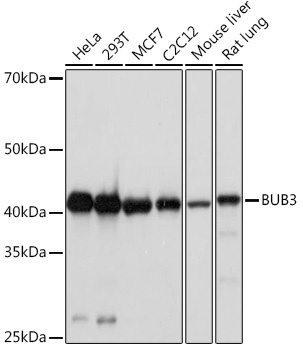
Western blot analysis of various lysates using BUB3 Rabbit mAb (CAB8831) at 1:1000 dilution. Secondary antibody: HRP-conjugated Goat anti-Rabbit IgG (H+L) (CABS014) at 1:10000 dilution. Lysates/proteins: 25μg per lane. Blocking buffer: 3% nonfat dry milk in TBST. Detection: ECL Basic Kit (AbGn00020). Exposure time: 90s.
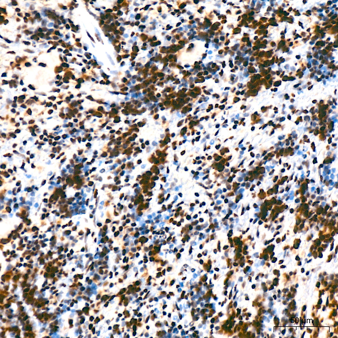
Immunohistochemistry analysis of paraffin-embedded Rat spleen tissue using BUB3 Rabbit mAb (CAB8831) at a dilution of 1:200 (40x lens). High pressure antigen retrieval was performed with 0.01 M citrate buffer (pH 6.0) prior to IHC staining.
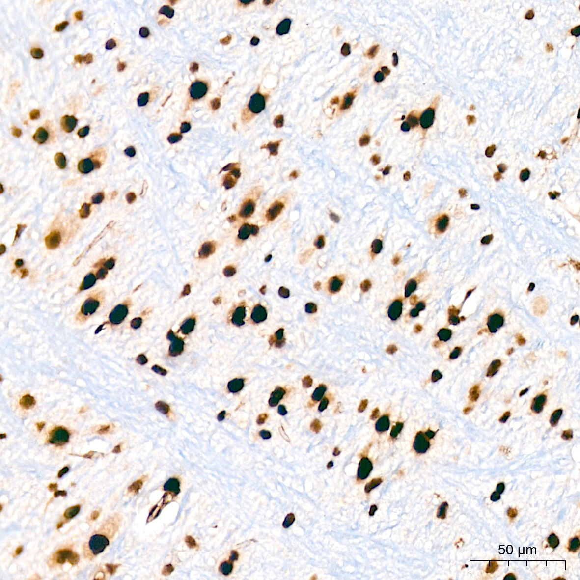
Immunohistochemistry analysis of paraffin-embedded Mouse brain tissue using BUB3 Rabbit mAb (CAB8831) at a dilution of 1:200 (40x lens). High pressure antigen retrieval was performed with 0.01 M citrate buffer (pH 6.0) prior to IHC staining.
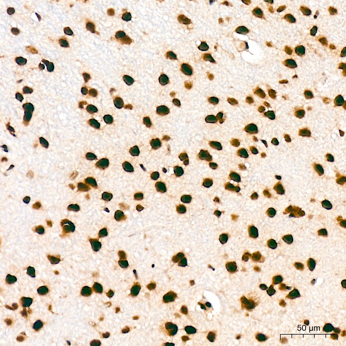
Immunohistochemistry analysis of paraffin-embedded Rat brain tissue using BUB3 Rabbit mAb (CAB8831) at a dilution of 1:200 (40x lens). High pressure antigen retrieval was performed with 0.01 M citrate buffer (pH 6.0) prior to IHC staining.
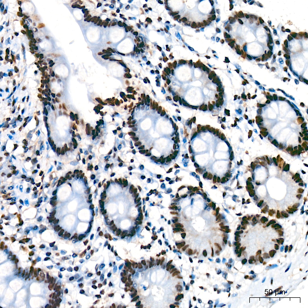
Immunohistochemistry analysis of paraffin-embedded Human colon tissue using BUB3 Rabbit mAb (CAB8831) at a dilution of 1:200 (40x lens). High pressure antigen retrieval was performed with 0.01 M citrate buffer (pH 6.0) prior to IHC staining.
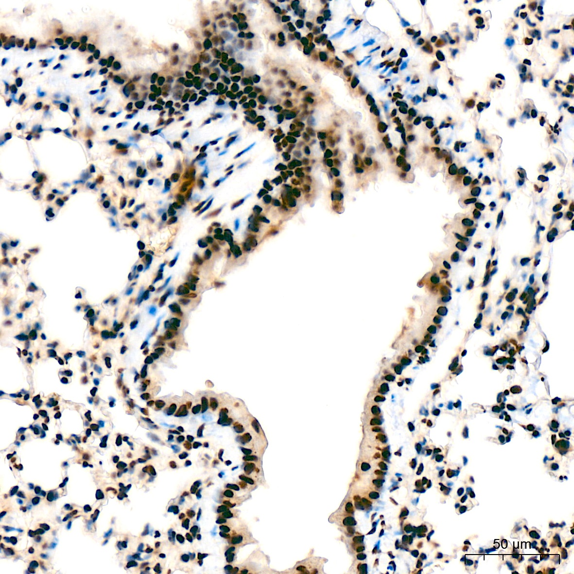
Immunohistochemistry analysis of paraffin-embedded Mouse lung tissue using BUB3 Rabbit mAb (CAB8831) at a dilution of 1:200 (40x lens). High pressure antigen retrieval was performed with 0.01 M citrate buffer (pH 6.0) prior to IHC staining.
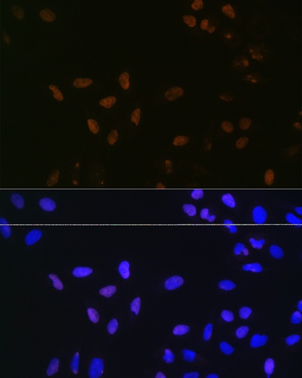
Immunofluorescence analysis of U-2 OS cells using BUB3 Rabbit mAb (CAB8831) at dilution of 1:100 (40x lens). Secondary antibody: Cy3-conjugated Goat anti-Rabbit IgG (H+L) (CABS007) at 1:500 dilution. Blue: DAPI for nuclear staining.