The C4D Monoclonal Antibody (CAB3545) is a high-quality antibody developed for reliable detection and analysis of target proteins. This antibody, raised in rabbits, demonstrates high reactivity with human samples and is validated for use in various applications such as Western blot and immunohistochemistry.Complement C4A is a critical player in the immune system, aiding in the recognition and clearance of pathogens and damaged cells.
This antibody is validated for use in WB, ELISA, IF-P applications and has demonstrated reactivity against Human samples.
Product Name:
C4D Monoclonal Antibody
SKU:
CAB3545
Size:
20μL, 100μL
Reactivity:
Human
Clone Number:
ARC2037
Conjugate:
Unconjugated
Immunogen:
Recombinant protein (or fragment).This information is considered to be commercially sensitive.
This gene encodes the acidic form of complement factor 4, part of the classical activation pathway. The protein is expressed as a single chain precursor which is proteolytically cleaved into a trimer of alpha, beta, and gamma chains prior to secretion. The trimer provides a surface for interaction between the antigen-antibody complex and other complement components. The alpha chain is cleaved to release C4 anaphylatoxin, an antimicrobial peptide and a mediator of local inflammation. Deficiency of this protein is associated with systemic lupus erythematosus and type I diabetes mellitus. This gene localizes to the major histocompatibility complex (MHC) class III region on chromosome 6. Varying haplotypes of this gene cluster exist, such that individuals may have 1, 2, or 3 copies of this gene. Two transcript variants encoding different isoforms have been found for this gene.
Purification Method
Affinity purification
Gene ID
720
Buffer Information
Store at -20℃. Avoid freeze / thaw cycles. Buffer: PBS containing 50% glycerol and 0.05% BSA, preserved with proclin300 or sodium azide, pH 7.3.
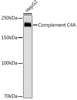
Western blot analysis of lysates from HepG2 cells, using Complement C4A Rabbit mAb (CAB3545) at 1:1000 dilution. Secondary antibody: HRP-conjugated Goat anti-Rabbit IgG (H+L) (CABS014) at 1:10000 dilution. Lysates/proteins: 25μg per lane. Blocking buffer: 3% nonfat dry milk in TBST. Detection: ECL Basic Kit (AbGn00020). Exposure time: 3s.
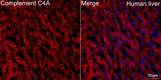
Confocal imaging of paraffin-embedded Human liver tissue using Complement C4A Rabbit mAb (CAB3545, dilution 1:100) followed by a further incubation with Cy3 Goat Anti-Rabbit IgG (H+L) (CABS007, dilution 1:500) (Red). DAPI was used for nuclear staining (Blue). Objective: 40x. Perform high pressure antigen retrieval with 0.01 M citrate buffer (pH 6.0) prior to IF staining.