The CAMLG Antibody (CAB13720) is a high-quality antibody developed for reliable detection and analysis of target proteins. This antibody, produced in rabbits, is highly specific for detecting CAMLG in human samples and is suitable for use in Western blot applications. By binding to CAMLG, researchers can study its function and expression levels in various cell types, aiding in investigations related to immune system regulation and potentially leading to insights into disease mechanisms.CAMLG, also known as calcium signal-modulating cyclophilin ligand, is known to play a role in calcium signaling and immune responses, making it a promising target for research on autoimmune diseases, inflammation, and cancer.
This antibody is validated for use in WB, IHC-P, IF/ICC, ELISA applications and has demonstrated reactivity against Human, Mouse, Rat samples.
Product Name:
CAMLG Antibody
SKU:
CAB13720
Size:
20μL, 100μL
Reactivity:
Human, Mouse, Rat
Conjugate:
Unconjugated
Immunogen:
Recombinant protein (or fragment).This information is considered to be commercially sensitive.
Recommended starting concentration is 1 μg/mL. Please optimize the concentration based on your specific assay requirements.
Synonyms:
CAML, GET2, CDG2Z, CAMLG
Positive Sample:
U-87MG, HepG2, HT-29, Mouse thymus, Mouse testis, Rat brain, Rat thymus
Cellular Localization:
Membrane, Multi-Pass Membrane Protein.
Calculated MW:
33kDa
Observed MW:
38kDa
The immunosuppressant drug cyclosporin A blocks a calcium-dependent signal from the T-cell receptor (TCR) that normally leads to T-cell activation. When bound to cyclophilin B, cyclosporin A binds and inactivates the key signaling intermediate calcineurin. The protein encoded by this gene functions similarly to cyclosporin A, binding to cyclophilin B and acting downstream of the TCR and upstream of calcineurin by causing an influx of calcium. This integral membrane protein appears to be a new participant in the calcium signal transduction pathway, implicating cyclophilin B in calcium signaling, even in the absence of cyclosporin.
Purification Method
Affinity purification
Gene ID
819
RRID
AB_2760580
Buffer Information
Store at -20℃. Avoid freeze / thaw cycles. Buffer: PBS with 0.01% thimerosal,50% glycerol,pH7.3.
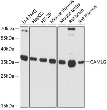
Western blot analysis of various lysates using CAMLG Rabbit pAb (CAB13720) at 1:3000 dilution. Secondary antibody: HRP-conjugated Goat anti-Rabbit IgG (H+L) (CABS014) at 1:10000 dilution. Lysates/proteins: 25μg per lane. Blocking buffer: 3% nonfat dry milk in TBST. Detection: ECL Basic Kit (AbGn00020). Exposure time: 90s.
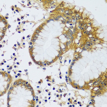
Immunohistochemistry analysis of paraffin-embedded Human stomach using CAMLG Rabbit pAb (CAB13720) at dilution of 1:100 (40x lens). Microwave antigen retrieval performed with 0.01M PBS Buffer (pH 7.2) prior to IHC staining.
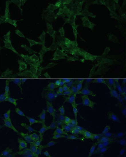
Immunofluorescence analysis of NIH-3T3 cells using CAMLG Rabbit pAb (CAB13720) at dilution of 1:100 (40x lens). Secondary antibody: Cy3-conjugated Goat anti-Rabbit IgG (H+L) (CABS007) at 1:500 dilution. Blue: DAPI for nuclear staining.
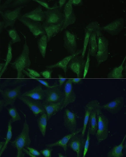
Immunofluorescence analysis of U-2 OS cells using CAMLG Rabbit pAb (CAB13720) at dilution of 1:100 (40x lens). Secondary antibody: Cy3-conjugated Goat anti-Rabbit IgG (H+L) (CABS007) at 1:500 dilution. Blue: DAPI for nuclear staining.