The Calpain 1 Antibody (CAB1172) is a high-quality antibody developed for reliable detection and analysis of target proteins. This antibody, raised in rabbits, is highly reactive with human samples and is validated for use in Western blot applications. It binds specifically to the CAPN1 protein, allowing for accurate detection and analysis in a variety of cell types, making it an ideal tool for studies in molecular biology and cell signaling pathways.
This antibody is validated for use in WB, IHC-P, IF/ICC, ELISA applications and has demonstrated reactivity against Human, Mouse, Rat samples.
Product Name:
Calpain 1 Antibody
SKU:
CAB1172
Size:
20μL, 100μL
Reactivity:
Human, Mouse, Rat
Conjugate:
Unconjugated
Immunogen:
Recombinant protein (or fragment).This information is considered to be commercially sensitive.
The calpains, calcium-activated neutral proteases, are nonlysosomal, intracellular cysteine proteases. The mammalian calpains include ubiquitous, stomach-specific, and muscle-specific proteins. The ubiquitous enzymes consist of heterodimers with distinct large, catalytic subunits associated with a common small, regulatory subunit. This gene encodes the large subunit of the ubiquitous enzyme, calpain 1. Several transcript variants encoding two different isoforms have been found for this gene.
Purification Method
Affinity purification
Gene ID
823
RRID
AB_2758715
Buffer Information
Store at -20℃. Avoid freeze / thaw cycles. Buffer: PBS with 0.09% Sodium azide,50% glycerol,pH7.3.
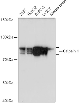
Western blot analysis of various lysates using Calpain 1 Rabbit pAb (CAB1172) at 1:1000 dilution. Secondary antibody: HRP-conjugated Goat anti-Rabbit IgG (H+L) (CABS014) at 1:10000 dilution. Lysates/proteins: 25μg per lane. Blocking buffer: 3% nonfat dry milk in TBST. Detection: ECL Basic Kit (AbGn00020). Exposure time: 90s.
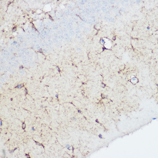
Immunohistochemistry analysis of paraffin-embedded Rat brain using Calpain 1 Rabbit pAb (CAB1172) at dilution of 1:100 (40x lens). High pressure antigen retrieval performed with 0.01M Citrate buffer (pH 6.0) prior to IHC staining.
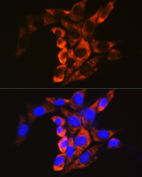
Immunofluorescence analysis of NIH/3T3 cells using Calpain 1 Rabbit pAb (CAB1172) at dilution of 1:100. Secondary antibody: Cy3-conjugated Goat anti-Rabbit IgG (H+L) (CABS007) at 1:500 dilution. Blue: DAPI for nuclear staining.