The CAPN7 Antibody (CAB17666) is a high-quality antibody developed for reliable detection and analysis of target proteins. This antibody, generated in rabbits, is highly specific for detecting CAPN7 in human samples and is validated for use in Western blot applications. By binding to the CAPN7 protein, this antibody allows for precise detection and analysis in various cell types, making it ideal for studies in molecular biology and cell biology research.CAPN7 is known for its involvement in cellular processes such as protein degradation, cytoskeletal remodeling, and signal transduction. Its role in regulating cell proliferation, differentiation, and apoptosis highlights its significance in various physiological and pathological conditions.
This antibody is validated for use in WB, IF/ICC, ELISA applications and has demonstrated reactivity against Human, Mouse, Rat samples.
Product Name:
CAPN7 Antibody
SKU:
CAB17666
Size:
20μL, 100μL
Reactivity:
Human, Mouse, Rat
Conjugate:
Unconjugated
Immunogen:
Recombinant protein (or fragment).This information is considered to be commercially sensitive.
Calpains are ubiquitous, well-conserved family of calcium-dependent, cysteine proteases. The calpain proteins are heterodimers consisting of an invariant small subunit and variable large subunits. The large subunit possesses a cysteine protease domain, and both subunits possess calcium-binding domains. Calpains have been implicated in neurodegenerative processes, as their activation can be triggered by calcium influx and oxidative stress. The function of the protein encoded by this gene is not known. An orthologue has been found in mouse but it seems to diverge from other family members. The mouse orthologue is thought to be calcium independent with protease activity.
Purification Method
Affinity purification
Gene ID
23473
RRID
AB_2768708
Buffer Information
Store at -20℃. Avoid freeze / thaw cycles. Buffer: PBS with 0.01% thimerosal,50% glycerol,pH7.3.
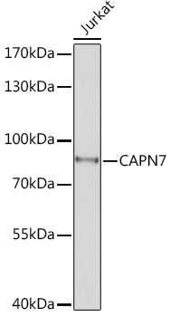
Western blot analysis of lysates from Jurkat cells, using CAPN7 Rabbit pAb (CAB17666) at 1:1000 dilution. Secondary antibody: HRP-conjugated Goat anti-Rabbit IgG (H+L) (CABS014) at 1:10000 dilution. Lysates/proteins: 25μg per lane. Blocking buffer: 3% nonfat dry milk in TBST. Detection: ECL Basic Kit (AbGn00020). Exposure time: 30s.
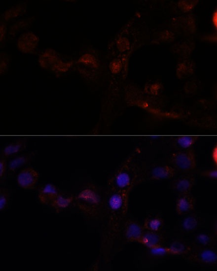
Immunofluorescence analysis of A431 cells using CAPN7 Rabbit pAb (CAB17666) at dilution of 1:100. Secondary antibody: Cy3-conjugated Goat anti-Rabbit IgG (H+L) (CABS007) at 1:500 dilution. Blue: DAPI for nuclear staining.