The Cathepsin G Monoclonal Antibody (CAB22048) is a high-quality antibody developed for reliable detection and analysis of target proteins. The protein encoded by this gene, a member of the peptidase S1 protein family, is found in azurophil granules of neutrophilic polymorphonuclear leukocytes. The encoded protease has a specificity similar to that of chymotrypsin C, and may participate in the killing and digestion of engulfed pathogens, and in connective tissue remodeling at sites of inflammation. In addition, the encoded protein is antimicrobial, with bacteriocidal activity against S. aureus and N. gonorrhoeae. Transcript variants utilizing alternative polyadenylation signals exist for this gene.
This antibody is validated for use in WB, ELISA applications and has demonstrated reactivity against Human samples.
Product Name:
Cathepsin G Monoclonal Antibody
SKU:
CAB22048
Size:
100μL, 20μL
Reactivity:
Human
Clone Number:
ARC54792
Conjugate:
Unconjugated
Immunogen:
Synthetic peptide. This information is considered to be commercially sensitive.
Tested Applications:
WBELISA
Recommended Dilution:
WB
1:500 - 1:1000
ELISA
Recommended starting concentration is 1 μg/mL. Please optimize the concentration based on your specific assay requirements.
Synonyms:
CG, CATG, Cathepsin G
Positive Sample:
THP-1
Cellular Localization:
Cell Surface.
Calculated MW:
29kDa
Observed MW:
29kDa
The protein encoded by this gene, a member of the peptidase S1 protein family, is found in azurophil granules of neutrophilic polymorphonuclear leukocytes. The encoded protease has a specificity similar to that of chymotrypsin C, and may participate in the killing and digestion of engulfed pathogens, and in connective tissue remodeling at sites of inflammation. In addition, the encoded protein is antimicrobial, with bacteriocidal activity against S. aureus and N. gonorrhoeae. Transcript variants utilizing alternative polyadenylation signals exist for this gene.
Purification Method
Affinity purification
Gene ID
1511
Buffer Information
Store at -20℃. Avoid freeze / thaw cycles. Buffer: PBS containing 50% glycerol and 0.05% BSA, preserved with proclin300 or sodium azide, pH 7.3.
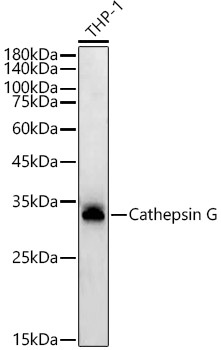
Western blot analysis of lysates from THP-1 cells, using Cathepsin G Rabbit mAb (CAB22048) at1:1000 dilution. Secondary antibody: HRP-conjugated Goat anti-Rabbit IgG (H+L) (AS014) at 1:10000 dilution. Lysates/proteins: 25μg per lane. Blocking buffer: 3% nonfat dry milk in TBST. Detection: ECL Basic Kit (AbGn00020). Exposure time: 60s.
 at1:1000 dilution. Secondary antibody: HRP Goat Anti-Rabbit IgG (H+L) at 1:10000 dilution. Lysates/proteins: 25μg per lane. Blocking buffer: 3% nonfat dry milk in TBST.")
 at1:1000 dilution. Secondary antibody: HRP Goat Anti-Rabbit IgG (H+L) at 1:10000 dilution. Lysates/proteins: 25μg per lane. Blocking buffer: 3% nonfat dry milk in TBST.")