The AAK1 Polyclonal Antibody (CAB22105) is a high-quality antibody developed for reliable detection and analysis of target proteins. This antibody, generated in rabbits, is highly specific for human samples and is validated for use in Western blot applications. By targeting the AAK1 protein, this antibody allows for precise detection and analysis in a variety of cell types, making it ideal for investigations in oncology and drug development. AAK1, also known as AP2 associated protein kinase 1, plays a crucial role in controlling cell growth, proliferation, and survival, making it a promising target for therapeutic intervention.
This antibody is validated for use in WB, ELISA applications and has demonstrated reactivity against Human samples.
Product Name:
AAK1 Polyclonal Antibody
SKU:
CAB22105
Size:
20μL, 100μL
Reactivity:
Human
Conjugate:
Unconjugated
Immunogen:
Synthetic peptide. This information is considered to be commercially sensitive.
This gene encodes a member of the SNF1 subfamily of serine/threonine protein kinases. Adaptor-related protein complex 2 (AP-2 complexes) functions during receptor-mediated endocytosis to trigger clathrin assembly, interact with membrane-bound receptors, and recruit encodytic accessory factors. The encoded protein interacts with and phosphorylates a subunit of the AP-2 complex, which promotes binding of AP-2 to sorting signals found in membrane-bound receptors and subsequent receptor endocytosis. Its kinase activity is stimulated by clathrin. This kinase has been shown to play an important role in regulating the clathrin-mediated endocytosis of the rabies virus, facilitating infection. Inhibitors of this kinase are being studied as candidate therapeutics to disrupt the entry of viruses, including SARS-CoV-2, into target cells. It is also involved in positive regulation of Notch pathway signaling in mammals. Alternatively spliced transcript variants have been described, but their biological validity has not been determined.
Purification Method
Affinity purification
Gene ID
22848
Buffer Information
Store at -20℃. Avoid freeze / thaw cycles. Buffer: PBS containing 50% glycerol, preserved with proclin300 or sodium azide, pH 7.3.
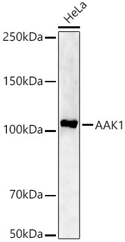
Western blot analysis of lysates from HeLa cells, using AAK1 Rabbit pAb (CAB22105) at 1:1000 dilution. Secondary antibody: HRP-conjugated Goat anti-Rabbit IgG (H+L) (CABS014) at 1:10000 dilution. Lysates/proteins: 25μg per lane. Blocking buffer: 3% nonfat dry milk in TBST. Detection: ECL Enhanced Kit (AbGn00021). Exposure time: 90s.
 at 1:1000 dilution. Secondary antibody: HRP Goat Anti-Rabbit IgG (H+L) at 1:10000 dilution. Lysates/proteins: 25ug per lane. Blocking buffer: 3% nonfat dry milk in TBST.")
 at 1:1000 dilution. Secondary antibody: HRP Goat Anti-Rabbit IgG (H+L) at 1:10000 dilution. Lysates/proteins: 25ug per lane. Blocking buffer: 3% nonfat dry milk in TBST.")


