The CD31/PECAM1 Monoclonal Antibody (CAB19014) is a high-quality antibody developed for reliable detection and analysis of target proteins. The antibody, developed using rabbit monoclonal technology, exhibits high specificity and sensitivity for human samples and is optimized for various applications including immunofluorescence, immunohistochemistry, and flow cytometry.CD31 is a key player in vascular biology, regulating processes such as leukocyte migration, angiogenesis, and platelet activation. Its role in mediating cell-cell adhesion and transendothelial migration makes it a crucial target for research in cardiovascular diseases, inflammation, and cancer metastasis.
This antibody is validated for use in WB, IHC-P, ELISA, IF-F, IF-P applications and has demonstrated reactivity against Human, Mouse, Rat samples.
Product Name:
CD31/PECAM1 Monoclonal Antibody
SKU:
CAB19014
Size:
20μL, 100μL
Reactivity:
Human, Mouse, Rat
Clone Number:
ARC50362
Conjugate:
Unconjugated
Immunogen:
Recombinant protein (or fragment).This information is considered to be commercially sensitive.
Cell Junction, Cell Junction, Cell Membrane, Lipid-Anchor, Single-Pass Type I Membrane Protein.
Calculated MW:
83kDa
Observed MW:
135kDa
The protein encoded by this gene is found on the surface of platelets, monocytes, neutrophils, and some types of T-cells, and makes up a large portion of endothelial cell intercellular junctions. The encoded protein is a member of the immunoglobulin superfamily and is likely involved in leukocyte migration, angiogenesis, and integrin activation.
Purification Method
Affinity purification
Gene ID
5175
RRID
AB_2862506
Buffer Information
Store at -20℃. Avoid freeze / thaw cycles. Buffer: PBS with 0.09% Sodium azide,0.05% BSA,50% glycerol,pH7.3.
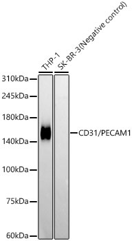
Western blot analysis of various lysates, using CD31/PECAM1 Rabbit mAb (CAB19014) at 1:12500 dilution. Secondary antibody: HRP-conjugated Goat anti-Rabbit IgG (H+L) (CABS014) at 1:10000 dilution. Lysates/proteins: 25μg per lane. Blocking buffer: 3% nonfat dry milk in TBST. Detection: ECL Basic Kit (AbGn00020). Negative control (NC): SK-BR-3 Exposure time: 180s.
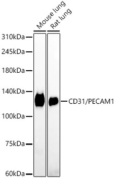
Western blot analysis of various lysates, using CD31/PECAM1 Rabbit mAb (CAB19014) at 1:12500 dilution. Secondary antibody: HRP-conjugated Goat anti-Rabbit IgG (H+L) (CABS014) at 1:10000 dilution. Lysates/proteins: 25μg per lane. Blocking buffer: 3% nonfat dry milk in TBST. Detection: ECL Enhanced Kit (AbGn00021). Exposure time: 180s.
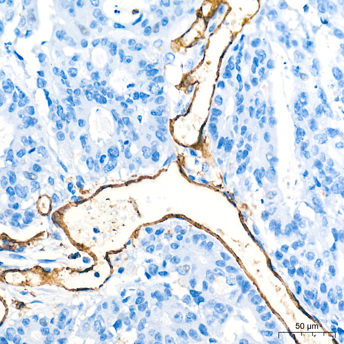
Immunohistochemistry analysis of paraffin-embedded Human colon carcinoma tissue using CD31/PECAM1 Rabbit mAb (CAB19014) at a dilution of 1:500 (40x lens). High pressure antigen retrieval performed with 0.01M Tris-EDTA Buffer (pH 9.0) prior to IHC staining.
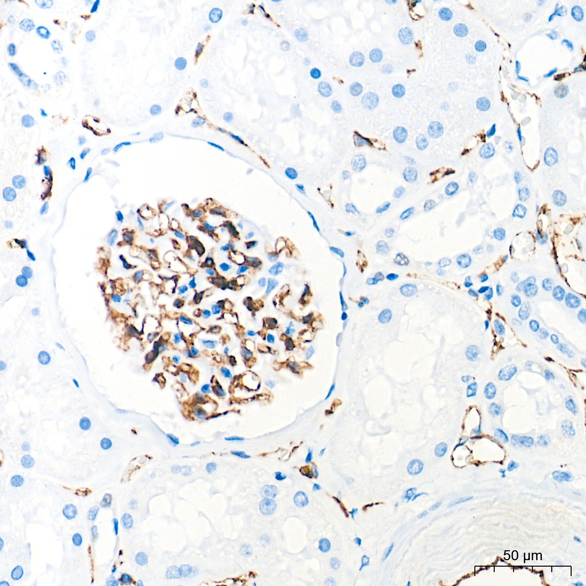
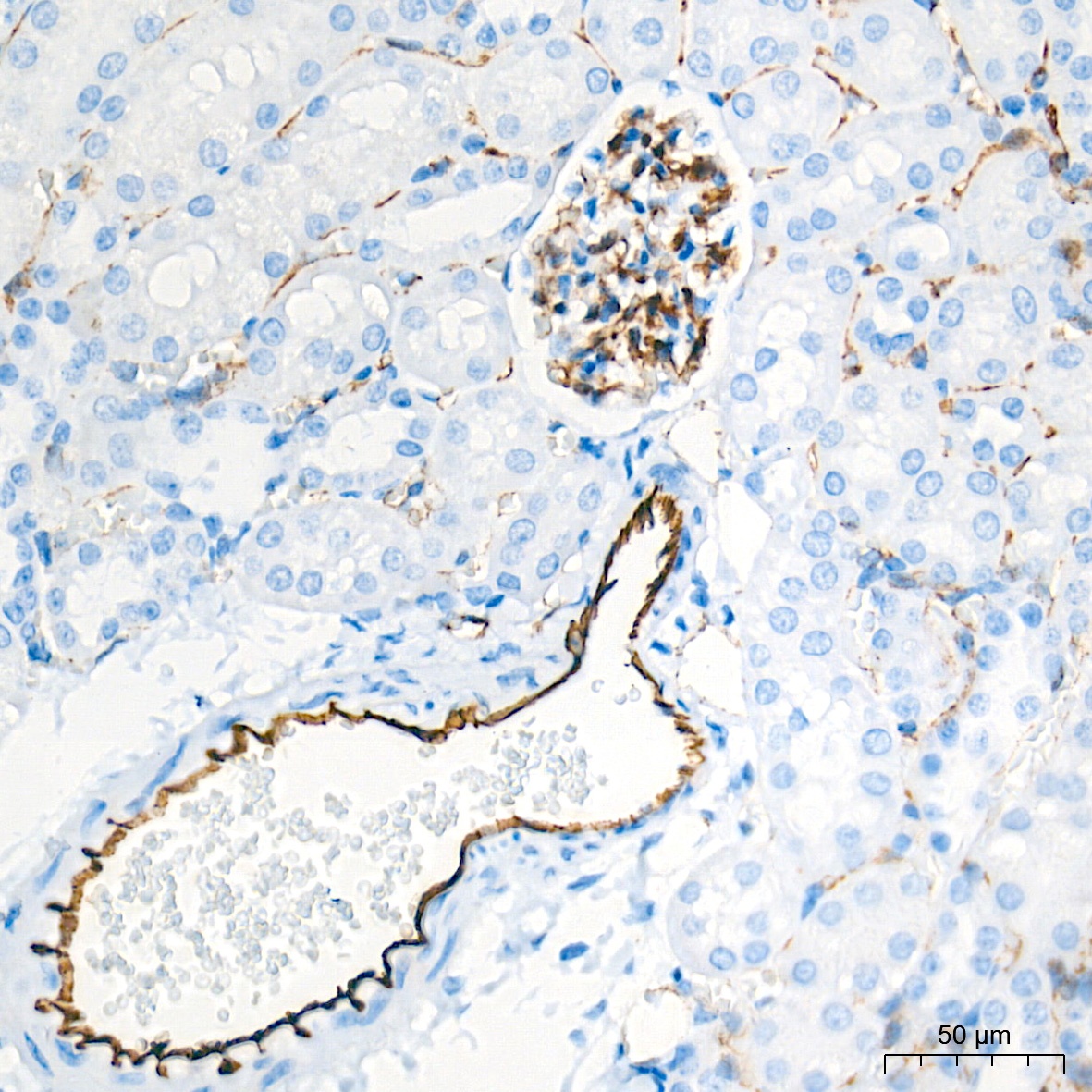
Immunohistochemistry analysis of paraffin-embedded Human kidney tissue using CD31/PECAM1 Rabbit mAb (CAB19014) at a dilution of 1:500 (40x lens). High pressure antigen retrieval performed with 0.01M Tris-EDTA Buffer (pH 9.0) prior to IHC staining.
Immunohistochemistry analysis of paraffin-embedded Mouse kidney tissue using CD31/PECAM1 Rabbit mAb (CAB19014) at a dilution of 1:500 (40x lens). High pressure antigen retrieval performed with 0.01M Tris-EDTA Buffer (pH 9.0) prior to IHC staining.
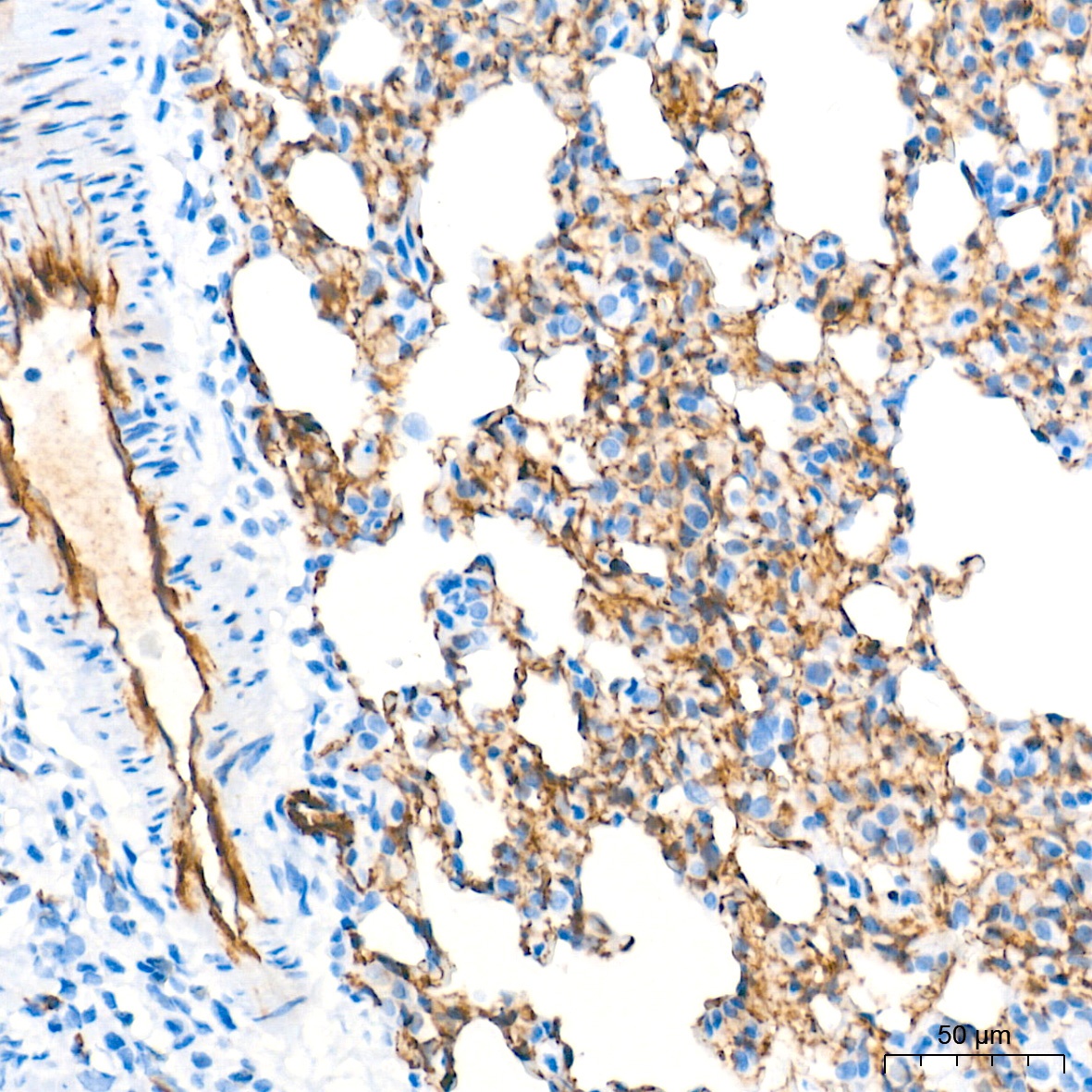
Immunohistochemistry analysis of paraffin-embedded Rat lung tissue using CD31/PECAM1 Rabbit mAb (CAB19014) at a dilution of 1:500 (40x lens). High pressure antigen retrieval performed with 0.01M Tris-EDTA Buffer (pH 9.0) prior to IHC staining.
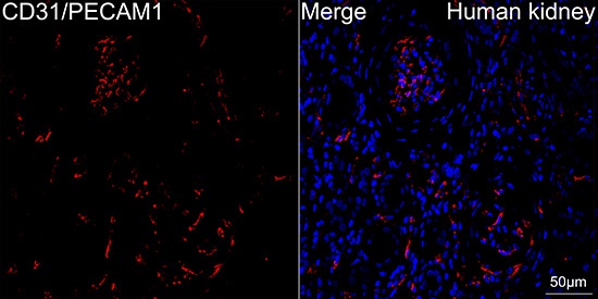
Confocal imaging of paraffin-embedded Human kidney tissue using CD31/PECAM1 Rabbit mAb (CAB19014, dilution 1:200) followed by a further incubation with Cy3 Goat Anti-Rabbit IgG (H+L) (CABS007, dilution 1:500) (Red). DAPI was used for nuclear staining (Blue). Objective: 40x.Perform high pressure antigen retrieval with 0.01M citrate buffer (pH 6.0) prior to IF staining.
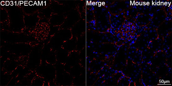
Confocal imaging of paraffin-embedded Mouse kidney tissue using CD31/PECAM1 Rabbit mAb (CAB19014, dilution 1:200) followed by a further incubation with Cy3 Goat Anti-Rabbit IgG (H+L) (CABS007, dilution 1:500) (Red). DAPI was used for nuclear staining (Blue). High pressure antigen retrieval performed with 0.01M Citrate Buffer (pH 6.0) prior to IF staining. Objective: 40x.
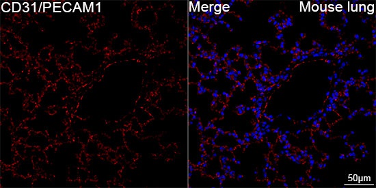
Confocal imaging of paraffin-embedded Mouse lung tissue using CD31/PECAM1 Rabbit mAb (CAB19014, dilution 1:200) followed by a further incubation with Cy3 Goat Anti-Rabbit IgG (H+L) (CABS007, dilution 1:500) (Red). DAPI was used for nuclear staining (Blue). High pressure antigen retrieval performed with 0.01M Citrate Buffer (pH 6.0) prior to IF staining. Objective: 40x.
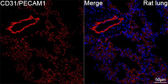
Confocal imaging of paraffin-embedded Rat lung tissue using CD31/PECAM1 Rabbit mAb (CAB19014, dilution 1:200) followed by a further incubation with Cy3 Goat Anti-Rabbit IgG (H+L) (CABS007, dilution 1:500) (Red). DAPI was used for nuclear staining (Blue). High pressure antigen retrieval performed with 0.01M Citrate Buffer (pH 6.0) prior to IF staining. Objective: 40x.


")