The CD3E Antibody (CAB1753) is a high-quality antibody developed for reliable detection and analysis of target proteins. This antibody, produced in rabbits, exhibits high specificity for human samples and is validated for use in Western blot applications. By binding to the CD3E protein, this antibody enables precise detection and analysis in a variety of cell types, making it suitable for investigations in immunology and cancer research.CD3E is essential for T-cell activation and function, playing a key role in immune surveillance and response to antigens.
This antibody is validated for use in WB, IHC-P, IF/ICC, IP, FC, ELISA, IF-P applications and has demonstrated reactivity against Human, Mouse, Rat samples.
Product Name:
CD3E Antibody
SKU:
CAB1753
Size:
20μL, 100μL
Reactivity:
Human, Mouse, Rat
Conjugate:
Unconjugated
Immunogen:
Recombinant protein (or fragment).This information is considered to be commercially sensitive.
0.5μg-4μg antibody for 200μg-400μg extracts of whole cells
IF/ICC
1:50 - 1:200
IF-P
1:50 - 1:200
IHC-P
1:50 - 1:200
FC
1:100 - 1:500
ELISA
Recommended starting concentration is 1 μg/mL. Please optimize the concentration based on your specific assay requirements.
Synonyms:
T3E, TCRE, IMD18, CD3epsilon, CD3E
Positive Sample:
Jurkat
Cellular Localization:
Cell Membrane, Single-Pass Type I Membrane Protein.
Calculated MW:
23kDa
Observed MW:
23kDa
The protein encoded by this gene is the CD3-epsilon polypeptide, which together with CD3-gamma, -delta and -zeta, and the T-cell receptor alpha/beta and gamma/delta heterodimers, forms the T-cell receptor-CD3 complex. This complex plays an important role in coupling antigen recognition to several intracellular signal-transduction pathways. The genes encoding the epsilon, gamma and delta polypeptides are located in the same cluster on chromosome 11. The epsilon polypeptide plays an essential role in T-cell development. Defects in this gene cause immunodeficiency. This gene has also been linked to a susceptibility to type I diabetes in women.
Purification Method
Affinity purification
Gene ID
916
RRID
AB_2763797
Buffer Information
Store at -20℃. Avoid freeze / thaw cycles. Buffer: PBS containing 50% glycerol, preserved with proclin300 or sodium azide, pH 7.3.
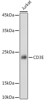
Western blot analysis of lysates from Jurkat cells, using CD3E Rabbit pAb (CAB1753) at 1:1000 dilution. Secondary antibody: HRP-conjugated Goat anti-Rabbit IgG (H+L) (CABS014) at 1:10000 dilution. Lysates/proteins: 25μg per lane. Blocking buffer: 3% nonfat dry milk in TBST. Detection: ECL Basic Kit (AbGn00020). Exposure time: 1s.
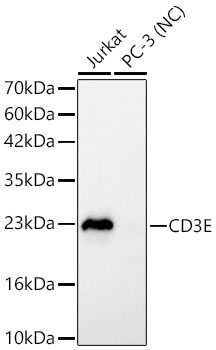
Western blot analysis of various lysates using Human CD3E Rabbit pAb (CAB1753) at 1:900 dilution. Secondary antibody: HRP-conjugated Goat anti-Rabbit IgG (H+L) (CABS014) at 1:10000 dilution. Lysates/proteins: 25 μg per lane. Blocking buffer: 3% nonfat dry milk in TBST. Detection: ECL Basic Kit (AbGn00020). Negative control (NC): PC-3. Exposure time: 1s.
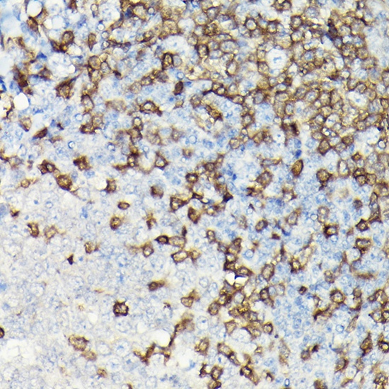
Immunohistochemistry analysis of paraffin-embedded Human tonsil using CD3E Rabbit pAb (CAB1753) at dilution of 1:200 (40x lens). High pressure antigen retrieval performed with 0.01M Citrate buffer (pH 6.0) prior to IHC staining.
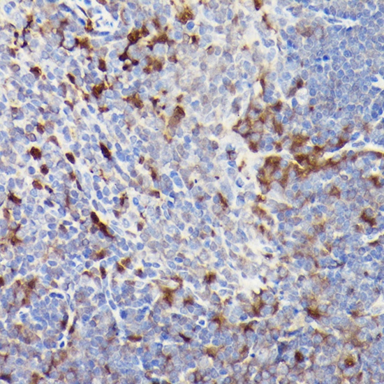
Immunohistochemistry analysis of paraffin-embedded Mouse spleen using CD3E Rabbit pAb (CAB1753) at dilution of 1:200 (40x lens). High pressure antigen retrieval performed with 0.01M Citrate buffer (pH 6.0) prior to IHC staining.
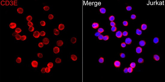
Immunofluorescence analysis of Jurkat cells using Human CD3E Rabbit pAb (CAB1753) at dilution of 1:100 (40x lens). Secondary antibody: Cy3-conjugated Goat anti-Rabbit IgG (H+L) (CABS007) at 1:500 dilution. Blue: DAPI for nuclear staining.
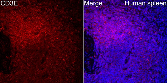
Immunofluorescence analysis of paraffin-embedded Human spleen tissue using CD3E Rabbit pAb (CAB1753) at a dilution of 1:100 (40x lens). Secondary antibody:Cy3 Goat Anti-Rabbit IgG (H+L) (CABS007) at 1:500 dilution. Blue: DAPI for nuclear staining. Perform high pressure antigen retrieval with 0.01 M citrate buffer (pH 6.0) prior to IF staining.
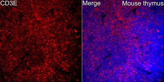
Immunofluorescence analysis of paraffin-embedded Mouse thyums tissue using CD3E Rabbit pAb(CAB1753) at a dilution of 1:100 (40x lens). Secondary antibody:Cy3 Goat Anti-Rabbit IgG (H+L) (CABS007) at 1:500 dilution. Blue: DAPI for nuclear staining. Perform high pressure antigen retrieval with 0.01 M citrate buffer (pH 6.0) prior to IF staining.
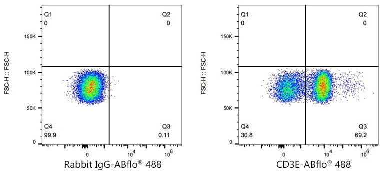
Flow cytometry: 1X10^6 Human PBMC were surface-stained with Rabbit IgG isotype control (AC042,2 μg/mL,left) or CD3E Rabbit pAb (CAB1753,2 μg/mL,right), followed by FITC conjugated goat anti-Rabbit pAb staining.