The CD6 Antibody (CAB8107) is a high-quality antibody developed for reliable detection and analysis of target proteins. This antibody, produced in rabbits, exhibits high reactivity with human samples and is validated for use in Western blot applications. By targeting the CD6 protein, this antibody enables precise detection and analysis in various cell types, making it ideal for investigations in immunology and cancer research.CD6, also known as T12 or TP120, is a crucial player in immune system regulation, acting as a co-stimulatory molecule that enhances T cell activation and proliferation.
This antibody is validated for use in WB, ELISA applications and has demonstrated reactivity against Human samples.
Product Name:
CD6 Antibody
SKU:
CAB8107
Size:
20μL, 100μL
Reactivity:
Human
Conjugate:
Unconjugated
Immunogen:
Recombinant protein (or fragment).This information is considered to be commercially sensitive.
Recommended starting concentration is 1 μg/mL. Please optimize the concentration based on your specific assay requirements.
Synonyms:
TP120, CD6
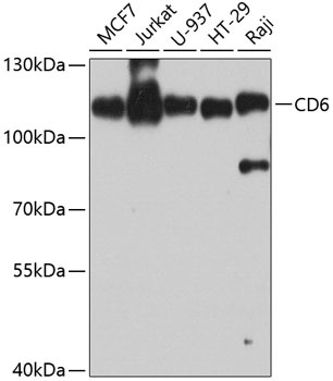
Positive Sample:
MCF7, Jurkat, U-937, HT-29, Raji
Cellular Localization:
Cell Membrane, Secreted, Single-Pass Type I Membrane Protein.
Calculated MW:
72kDa
Observed MW:
110kDa
This gene encodes a protein found on the outer membrane of T-lymphocytes as well as some other immune cells. The encoded protein contains three scavenger receptor cysteine-rich (SRCR) domains and a binding site for an activated leukocyte cell adhesion molecule. The gene product is important for continuation of T cell activation. This gene may be associated with susceptibility to multiple sclerosis (PMID: 19525953, 21849685). Multiple transcript variants encoding different isoforms have been found for this gene.
Purification Method
Affinity purification
Gene ID
923
RRID
AB_2768794
Buffer Information
Store at -20℃. Avoid freeze / thaw cycles. Buffer: PBS containing 50% glycerol, preserved with proclin300 or sodium azide, pH 7.3.
Western blot analysis of various lysates using CD6 Rabbit pAb (CAB8107) at 1:1000 dilution. Secondary antibody: HRP-conjugated Goat anti-Rabbit IgG (H+L) (CABS014) at 1:10000 dilution. Lysates/proteins: 25μg per lane. Blocking buffer: 3% nonfat dry milk in TBST. Detection: ECL Enhanced Kit (AbGn00021). Exposure time: 60s.