The CDA Antibody (CAB13959) is a high-quality antibody developed for reliable detection and analysis of target proteins. This antibody, derived from rabbit serum, is highly specific to human samples and has been validated for use in Western blot applications. By binding to the CD300A protein, this antibody enables researchers to detect and analyze CD300A in various cell types, making it ideal for studies in immunology and cancer research.CD300A, also known as an immune inhibitory receptor, plays a crucial role in maintaining immune homeostasis by regulating inflammation and inhibiting allergic reactions.
This antibody is validated for use in WB, IHC-P, IF/ICC, ELISA applications and has demonstrated reactivity against Human, Mouse samples.
Product Name:
CDA Antibody
SKU:
CAB13959
Size:
20μL, 100μL
Reactivity:
Human, Mouse
Conjugate:
Unconjugated
Immunogen:
Recombinant protein (or fragment).This information is considered to be commercially sensitive.
Recommended starting concentration is 1 μg/mL. Please optimize the concentration based on your specific assay requirements.
Synonyms:
CDD, CDA
Positive Sample:
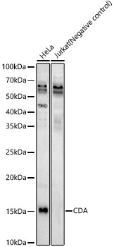
HeLa, Jurkat(Negative control), Mouse kidney
Cellular Localization:
Cytosol, Extracellular Region.
Calculated MW:
16kDa
Observed MW:
16kDa
This gene encodes an enzyme involved in pyrimidine salvaging. The encoded protein forms a homotetramer that catalyzes the irreversible hydrolytic deamination of cytidine and deoxycytidine to uridine and deoxyuridine, respectively. It is one of several deaminases responsible for maintaining the cellular pyrimidine pool. Mutations in this gene are associated with decreased sensitivity to the cytosine nucleoside analogue cytosine arabinoside used in the treatment of certain childhood leukemias.
Purification Method
Affinity purification
Gene ID
978
RRID
AB_2760813
Buffer Information
Store at -20℃. Avoid freeze / thaw cycles. Buffer: PBS containing 50% glycerol, preserved with proclin300 or sodium azide, pH 7.3.
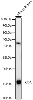
Western blot analysis of various lysates, using CDA Rabbit pAb (CAB13959) at 1:600 dilution. Secondary antibody: HRP-conjugated Goat anti-Rabbit IgG (H+L) (CABS014) at 1:10000 dilution. Lysates/proteins: 25μg per lane. Blocking buffer: 3% nonfat dry milk in TBST. Detection: ECL Basic Kit (AbGn00020). Exposure time: 90s.
Western blot analysis of various lysates, using CDA Rabbit pAb (CAB13959) at 1:600 dilution. Secondary antibody: HRP-conjugated Goat anti-Rabbit IgG (H+L) (CABS014) at 1:10000 dilution. Lysates/proteins: 25μg per lane. Blocking buffer: 3% nonfat dry milk in TBST. Detection: ECL Enhanced Kit (AbGn00021). Exposure time: 60s.
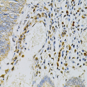
Immunohistochemistry analysis of paraffin-embedded Human colon carcinoma using CDA Rabbit pAb (CAB13959) at dilution of 1:100 (40x lens). Microwave antigen retrieval performed with 0.01M PBS Buffer (pH 7.2) prior to IHC staining.