The CDH11 Antibody (CAB12176) is a high-quality antibody developed for reliable detection and analysis of target proteins. This antibody, raised in rabbits, demonstrates high reactivity with human samples and has been validated for use in Western blot applications. By binding specifically to the Cadherin-11 protein, this antibody enables accurate detection and analysis in various cell types, making it an excellent choice for studies in developmental biology and cancer research.Cadherin-11, a member of the cadherin superfamily, plays a crucial role in various biological processes, including tissue morphogenesis and tumor metastasis.
This antibody is validated for use in WB, IF/ICC, ELISA applications and has demonstrated reactivity against Human, Mouse samples.
Product Name:
CDH11 Antibody
SKU:
CAB12176
Size:
20μL, 100μL
Reactivity:
Human, Mouse
Conjugate:
Unconjugated
Immunogen:
Recombinant protein (or fragment).This information is considered to be commercially sensitive.
Recommended starting concentration is 1 μg/mL. Please optimize the concentration based on your specific assay requirements.
Synonyms:
OB, ESWS, CAD11, CDHOB, OSF-4, TBHS2, CDH11
Positive Sample:
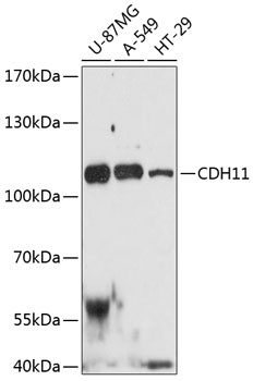
U-87MG, A-549, HT-29
Cellular Localization:
Cell Membrane, Single-Pass Type I Membrane Protein.
Calculated MW:
88kDa
Observed MW:
110kDa
This gene encodes a type II classical cadherin from the cadherin superfamily, integral membrane proteins that mediate calcium-dependent cell-cell adhesion. Mature cadherin proteins are composed of a large N-terminal extracellular domain, a single membrane-spanning domain, and a small, highly conserved C-terminal cytoplasmic domain. Type II (atypical) cadherins are defined based on their lack of a HAV cell adhesion recognition sequence specific to type I cadherins. Expression of this particular cadherin in osteoblastic cell lines, and its upregulation during differentiation, suggests a specific function in bone development and maintenance.
Purification Method
Affinity purification
Gene ID
1009
RRID
AB_2759063
Buffer Information
Store at -20℃. Avoid freeze / thaw cycles. Buffer: PBS with 0.01% thimerosal,50% glycerol,pH7.3.
Western blot analysis of various lysates using CDH11 Rabbit pAb (CAB12176) at 1:3000 dilution._Secondary antibody: HRP-conjugated Goat anti-Rabbit IgG (H+L) (CABS014) at 1:10000 dilution._Lysates/proteins: 25μg per lane._Blocking buffer: 3% nonfat dry milk in TBST._Detection: ECL Enhanced Kit (AbGn00021)._Exposure time: 90s.
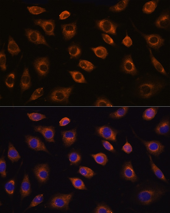
Immunofluorescence analysis of L929 cells using CDH11 Rabbit pAb (CAB12176) at dilution of 1:100. Secondary antibody: Cy3-conjugated Goat anti-Rabbit IgG (H+L) (CABS007) at 1:500 dilution. Blue: DAPI for nuclear staining.