The CRX Antibody (CAB5719) is a high-quality antibody developed for reliable detection and analysis of target proteins. This antibody, produced in rabbits, is highly specific to human samples, making it ideal for use in Western blot applications. By binding to the CRX protein, researchers can detect and analyze its expression in various cell types, enabling insights into its roles in vision and retinal development.
This antibody is validated for use in WB, ELISA, IF-P applications and has demonstrated reactivity against Mouse samples.
Product Name:
CRX Antibody
SKU:
CAB5719
Size:
20μL, 100μL
Reactivity:
Mouse
Conjugate:
Unconjugated
Immunogen:
Synthetic peptide. This information is considered to be commercially sensitive.
Recommended starting concentration is 1 μg/mL. Please optimize the concentration based on your specific assay requirements.
Synonyms:
CRD, LCA7, OTX3, CORD2, CRX
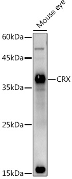
Positive Sample:
Mouse eye
Cellular Localization:
Nucleus.
Calculated MW:
32kDa
Observed MW:
37kDa
The protein encoded by this gene is a photoreceptor-specific transcription factor which plays a role in the differentiation of photoreceptor cells. This homeodomain protein is necessary for the maintenance of normal cone and rod function. Mutations in this gene are associated with photoreceptor degeneration, Leber congenital amaurosis type III and the autosomal dominant cone-rod dystrophy 2. Several alternatively spliced transcript variants of this gene have been described, but the full-length nature of some variants has not been determined.
Purification Method
Affinity purification
Gene ID
1406
RRID
AB_2766476
Buffer Information
Store at -20℃. Avoid freeze / thaw cycles. Buffer: PBS containing 50% glycerol, preserved with proclin300 or sodium azide, pH 7.3.
Western blot analysis of lysates from Mouse eye, using CRX Rabbit pAb (CAB5719) at 1:1000 dilution. Secondary antibody: HRP-conjugated Goat anti-Rabbit IgG (H+L) (CABS014) at 1:10000 dilution. Lysates/proteins: 25μg per lane. Blocking buffer: 3% nonfat dry milk in TBST. Detection: ECL Basic Kit (AbGn00020). Exposure time: 180s.
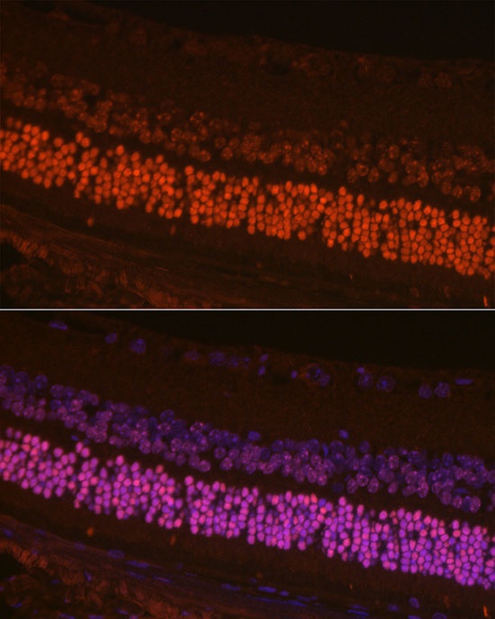
Immunofluorescence analysis of paraffin-embedded mouse retina using CRX Rabbit pAb (CAB5719) at dilution of 1:200 (40x lens). Secondary antibody: Cy3-conjugated Goat anti-Rabbit IgG (H+L) (CABS007) at 1:500 dilution. Blue: DAPI for nuclear staining.