The Cyclin A2 Monoclonal Antibody (CAB19036) is a high-quality antibody developed for reliable detection and analysis of target proteins. This antibody, generated in rabbits, specifically targets Cyclin A2, a key regulator of cell cycle progression and division. Validated for use in Western blot applications, this antibody allows for detection and analysis of Cyclin A2 levels in various cell types.Cyclin A2 plays a crucial role in controlling the transition from G1 to S phase during the cell cycle, making it a promising target for understanding the mechanisms underlying uncontrolled cell growth in cancer.
This antibody is validated for use in WB, IHC-P, IF/ICC, IP, ELISA applications and has demonstrated reactivity against Human, Mouse, Rat samples.
Product Name:
Cyclin A2 Monoclonal Antibody
SKU:
CAB19036
Size:
20μL, 100μL
Reactivity:
Human, Mouse, Rat
Clone Number:
ARC0359
Conjugate:
Unconjugated
Immunogen:
Recombinant protein (or fragment).This information is considered to be commercially sensitive.
0.5μg-4μg antibody for 200μg-400μg extracts of whole cells
ELISA
Recommended starting concentration is 1 μg/mL. Please optimize the concentration based on your specific assay requirements.
Synonyms:
CCN1, CCNA, Cyclin A2
Positive Sample:
HeLa, HCT 116, Jurkat, Mouse testis
Cellular Localization:
Cytoplasm, Nucleus.
Calculated MW:
49kDa
Observed MW:
55kDa
The protein encoded by this gene belongs to the highly conserved cyclin family, whose members function as regulators of the cell cycle. This protein binds and activates cyclin-dependent kinase 2 and thus promotes transition through G1/S and G2/M.
Purification Method
Affinity purification
Gene ID
890
RRID
AB_2862528
Buffer Information
Store at -20℃. Avoid freeze / thaw cycles. Buffer: PBS containing 50% glycerol and 0.05% BSA, preserved with proclin300 or sodium azide, pH 7.3.
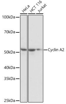
Western blot analysis of various lysates using Cyclin A2 Rabbit mAb (CAB19036) at 1:1000 dilution. Secondary antibody: HRP-conjugated Goat anti-Rabbit IgG (H+L) (CABS014) at 1:10000 dilution. Lysates/proteins: 25μg per lane. Blocking buffer: 3% nonfat dry milk in TBST. Detection: ECL Basic Kit (AbGn00020). Exposure time: 1s.
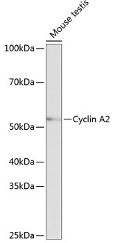
Western blot analysis of lysates from Mouse testis, using Cyclin A2 Rabbit mAb (CAB19036) at 1:1000 dilution. Secondary antibody: HRP-conjugated Goat anti-Rabbit IgG (H+L) (CABS014) at 1:10000 dilution. Lysates/proteins: 25μg per lane. Blocking buffer: 3% nonfat dry milk in TBST. Detection: ECL Basic Kit (AbGn00020). Exposure time: 10s.
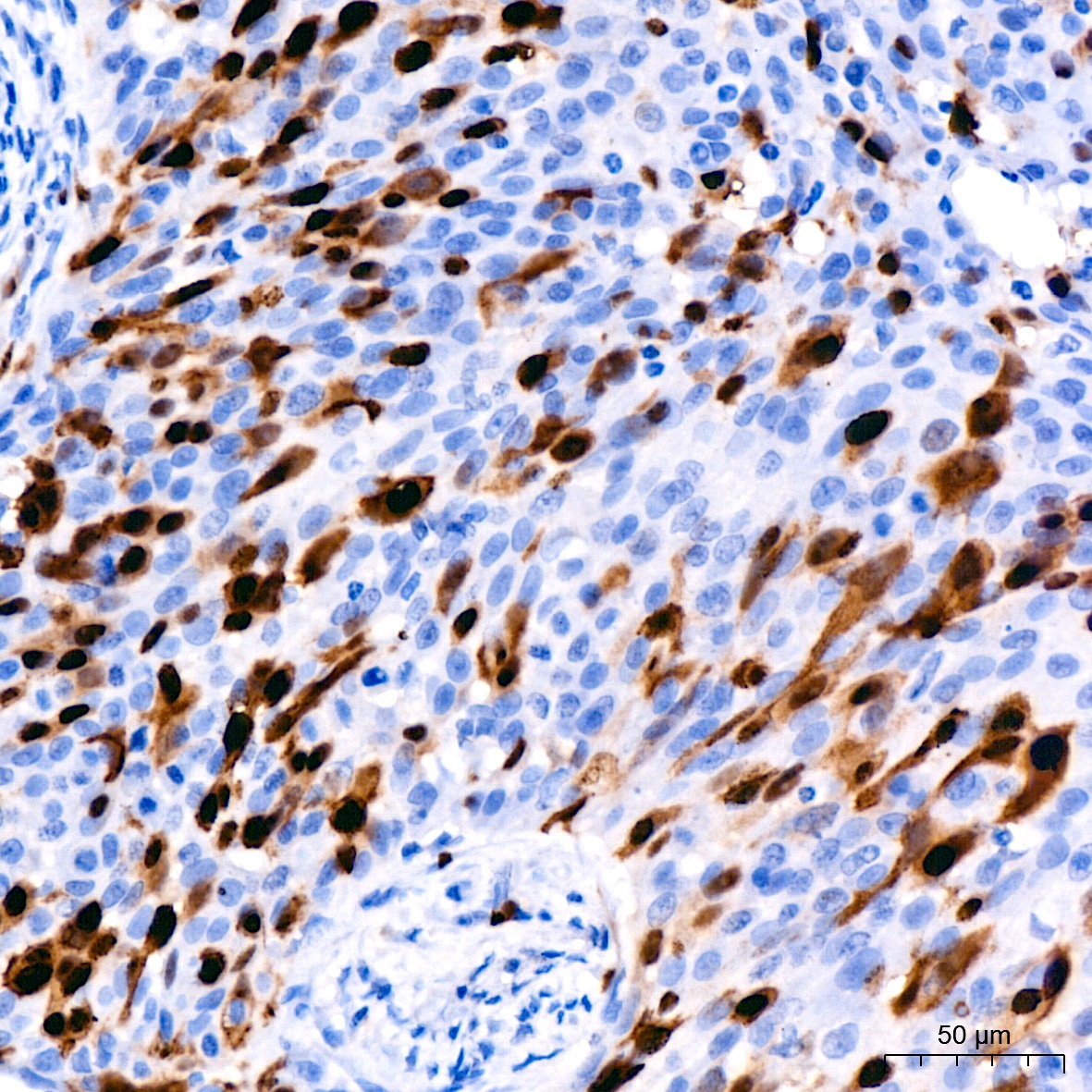
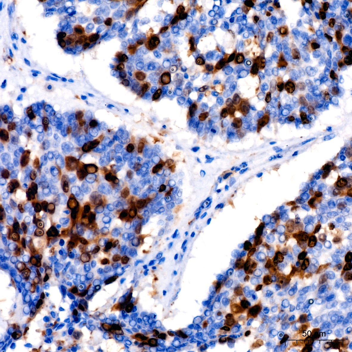
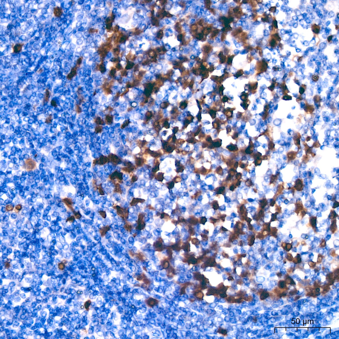
Immunohistochemistry analysis of paraffin-embedded Human cervical squamous cell carcinoma using Cyclin A2 Rabbit mAb (CAB19036) at dilution of 1:200 (40x lens). High pressure antigen retrieval performed with 0.01M Citrate buffer (pH 6.0) prior to IHC staining.
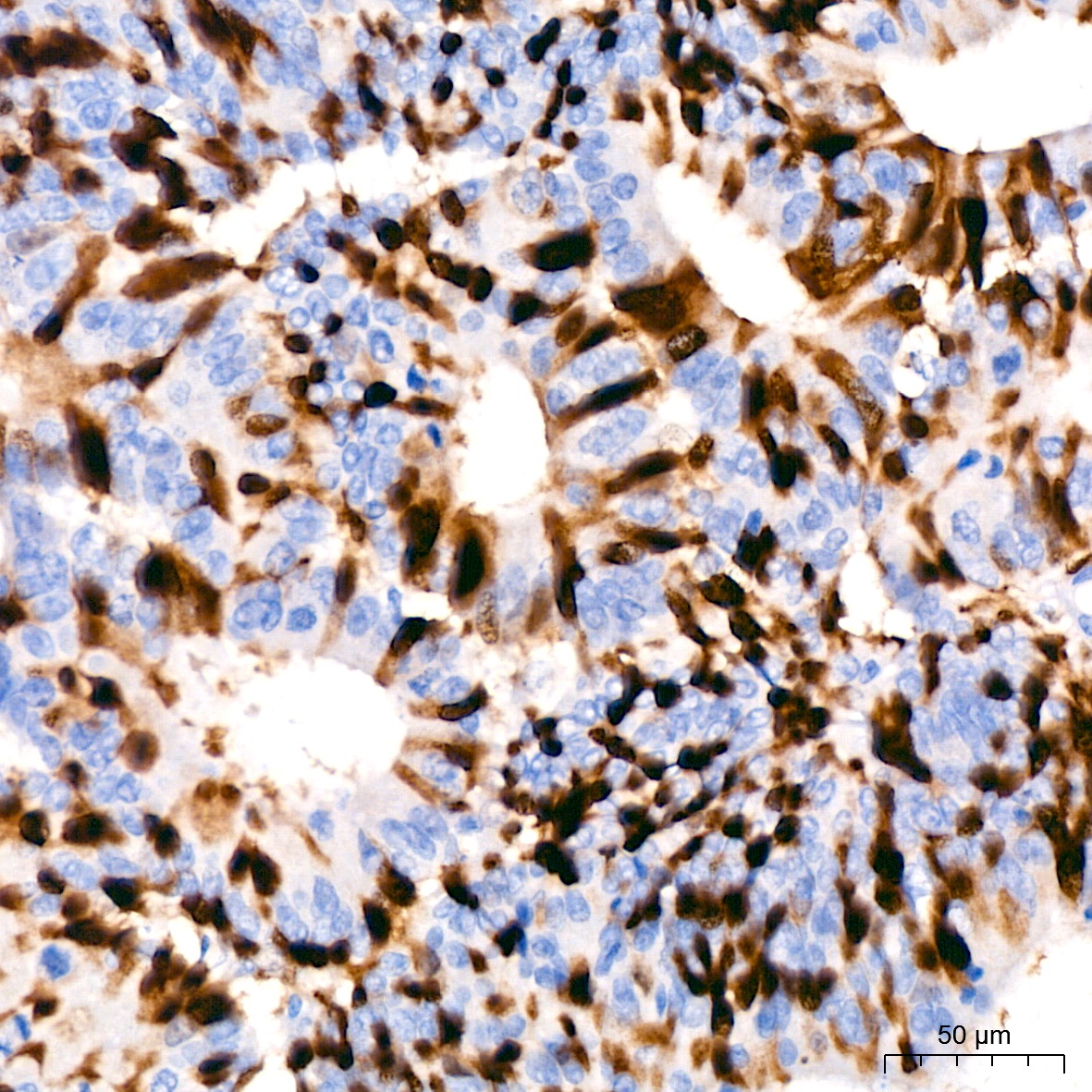
Immunohistochemistry analysis of paraffin-embedded Human colon carcinoma using Cyclin A2 Rabbit mAb (CAB19036) at dilution of 1:200 (40x lens). High pressure antigen retrieval performed with 0.01M Citrate buffer (pH 6.0) prior to IHC staining.
Immunohistochemistry analysis of paraffin-embedded Human lung cancer using Cyclin A2 Rabbit mAb (CAB19036) at dilution of 1:200 (40x lens). High pressure antigen retrieval performed with 0.01M Citrate buffer (pH 6.0) prior to IHC staining.
Immunohistochemistry analysis of paraffin-embedded Human tonsil using Cyclin A2 Rabbit mAb (CAB19036) at dilution of 1:200 (40x lens). High pressure antigen retrieval performed with 0.01M Citrate buffer (pH 6.0) prior to IHC staining.
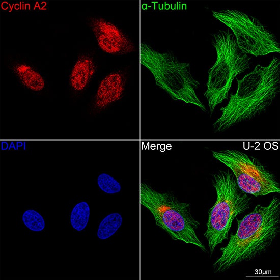
Confocal imaging of U-2 OS cells using Cyclin A2 Rabbit mAb (CAB19036,at dilution of 1:100) (Red). The cells were counterstained with α-Tubulin Mouse mAb (AC012,dilution 1:400) (Green). DAPI was used for nuclear staining (blue). Objective: 100x.
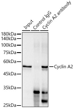
Immunoprecipitation analysis of 300 μg extracts of HeLa cells using 3 μg Cyclin A2 antibody (CAB19036). Western blot was performed from the immunoprecipitate using Cyclin A2 antibody (CAB19036) at a dilution of 1:1000.