The DOK1 Monoclonal Antibody (CAB19250) is a high-quality antibody developed for reliable detection and analysis of target proteins. This antibody, generated in rabbits, demonstrates high specificity and sensitivity when detecting DOK1 in human samples, making it an excellent choice for Western blotting experiments. By targeting the DOK1 protein, researchers can gain insights into its function and role in various cellular processes, particularly in the fields of immunology and cancer research.DOK1 is known for its involvement in cellular signaling pathways that control proliferation and differentiation, making it a potential target for therapeutic interventions in diseases like cancer and autoimmune disorders.
This antibody is validated for use in WB, IF/ICC, ELISA applications and has demonstrated reactivity against Human, Mouse samples.
Product Name:
DOK1 Monoclonal Antibody
SKU:
CAB19250
Size:
20μL, 100μL
Reactivity:
Human, Mouse
Clone Number:
ARC2410
Conjugate:
Unconjugated
Immunogen:
Synthetic peptide. This information is considered to be commercially sensitive.
Recommended starting concentration is 1 μg/mL. Please optimize the concentration based on your specific assay requirements.
Synonyms:
pp62, P62DOK, DOK1
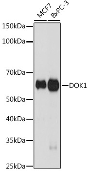
Positive Sample:
MCF7, BxPC-3
Cellular Localization:
Cytoplasm, Nucleus, Perinuclear Region.
Calculated MW:
52kDa
Observed MW:
62kDa
The protein encoded by this gene is part of a signal transduction pathway downstream of receptor tyrosine kinases. The encoded protein is a scaffold protein that helps form a platform for the assembly of multiprotein signaling complexes. Several transcript variants encoding different isoforms have been found for this gene.
Purification Method
Affinity purification
Gene ID
1796
Buffer Information
Store at -20℃. Avoid freeze / thaw cycles. Buffer: PBS containing 50% glycerol and 0.05% BSA, preserved with proclin300 or sodium azide, pH 7.3.
Western blot analysis of various lysates using DOK1 Rabbit mAb (CAB19250) at 1:1000 dilution. Secondary antibody: HRP-conjugated Goat anti-Rabbit IgG (H+L) (CABS014) at 1:10000 dilution. Lysates/proteins: 25μg per lane. Blocking buffer: 3% nonfat dry milk in TBST. Detection: ECL Basic Kit (AbGn00020). Exposure time: 90s.
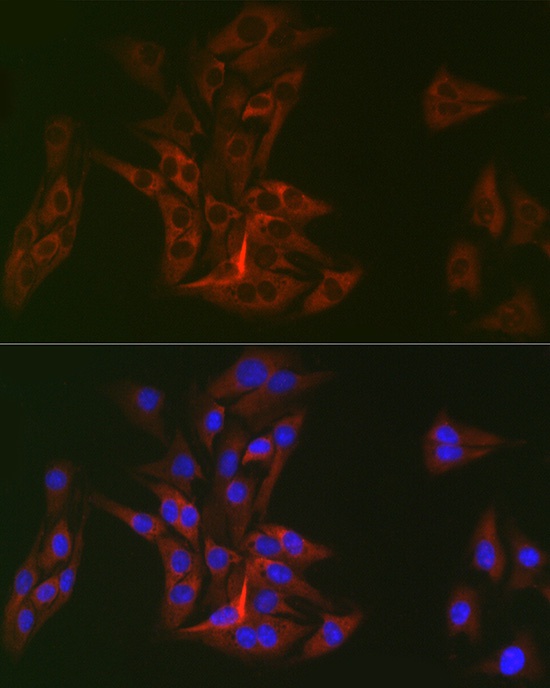
Immunofluorescence analysis of BALB-3T3 cells using DOK1 Rabbit mAb (CAB19250) at dilution of 1:100 (40x lens). Secondary antibody: Cy3-conjugated Goat anti-Rabbit IgG (H+L) (CABS007) at 1:500 dilution. Blue: DAPI for nuclear staining.